DALLAS – Jan. 24, 2018 – UT Southwestern Simmons Cancer Center scientists next month will begin testing a digital nanosensor that lights up cancer tissue to see whether it can improve the accuracy of cancer surgeries, thereby reducing cancer recurrence and surgical morbidity.
The nanosensor, which works by reacting to low pH, illuminates cancer like a lightbulb, sharply distinguishing cancerous tissue from healthy tissue, and making it easier for surgeons to remove cancer cells while leaving healthy, functional tissue intact.
“We synthesized an imaging probe that stays dark in normal tissues but switches on in solid tumors, behaving like a digital sensor with binary readouts between the two states,” said Dr. Jinming Gao, Professor of Pharmacology and Otolaryngology with the Harold C. Simmons Comprehensive Cancer Center at UT Southwestern Medical Center, which is recognizing its 75th anniversary this year. “Cancer is a diverse set of diseases, but it does have some universal features. As solid tumors ramp up, they eat more glucose and secrete lactic acid, so the microenvironment around the cancer cells is acidic.”
Dr. Gao and surgeon Dr. Baran Sumer, Associate Professor of Otolaryngology with the Simmons Cancer Center, developed the sensor, and have integrated it with a clinical camera that allows the surgeon to see the fluorescent areas. Their nanosensor not only lights up cancer, but suppresses the signal in normal tissue, leading to a sharp line between cancer and healthy tissue. It is expected to be effective in all solid tumors.
“This new digital nanosensor-guided surgery has several advantages for patients, including more accurate removal of tumors and greater preservation of normal tissue. These advantages can limit the extent of surgery and improve quality of life and, potentially, patient survival,” said Dr. Sumer, who leads the Head and Neck Oncology team at the Simmons Cancer Center.
First to be treated with the new technology will be breast cancer patients at the University of Groningen in the Netherlands, which is collaborating in the clinical trials. Patients will be intravenously injected with the nanosensor medication about 12 hours before surgery. Tumors will light up and remain fluorescent for one to two days. Dr. Sumer and Dr. Gao expect to have data on breast cancer and colon cancer surgeries by fall, then hope to move on to Phase II clinical trials in the U.S. and elsewhere.
“What we have is a digital probe that might eventually lead to more exact surgeries so that solid-cancer surgeries would not need to be followed up with radiation or chemotherapy,” said Dr. Sumer.
The nanosensor technology and the camera used to see the fluorescent cells are being developed by OncoNano Medicine, Inc., which was the recipient of a Cancer Prevention and Research Institute of Texas product development award to develop this technology. Dr. Gao and Dr. Sumer are paid consultants and scientific co-founders of OncoNano Medicine, Inc. UT Southwestern Medical Center has licensed the technology to OncoNano Medicine and has a financial interest in the research.
The Harold C. Simmons Comprehensive Cancer Center is the only NCI-designated Comprehensive Cancer Center in North Texas and one of just 49 NCI-designated Comprehensive Cancer Centers in the nation. Simmons Cancer Center is among just 30 U.S. cancer research centers to be designated by the NCI as a National Clinical Trials Network Lead Academic Participating Site.
###
About UT Southwestern Medical Center
UT Southwestern, one of the premier academic medical centers in the nation, integrates pioneering biomedical research with exceptional clinical care and education. The institution’s faculty has received six Nobel Prizes, and includes 22 members of the National Academy of Sciences, 18 members of the National Academy of Medicine, and 14 Howard Hughes Medical Institute Investigators. The faculty of more than 2,700 is responsible for groundbreaking medical advances and is committed to translating science-driven research quickly to new clinical treatments. UT Southwestern physicians provide care in about 80 specialties to more than 100,000 hospitalized patients, 600,000 emergency room cases, and oversee approximately 2.2 million outpatient visits a year.
A transistor-like pH nanoprobe for tumour detection and image-guided surgery
Tian Zhao, Gang Huang, Yang Li, Shunchun Yang, Saleh Ramezani, Zhiqiang Lin, Yiguang Wang, Xinpeng Ma, Zhiqun Zeng, Min Luo, Esther de Boer, Xian-Jin Xie, Joel Thibodeaux, Rolf A. Brekken, Xiankai Sun, Baran D. Sumer & Jinming Gao
Nature Biomedical Engineering 1, Article number: 0006 (2016)
doi:10.1038/s41551-016-0006
Download Citation
Biomedical engineering Cancer imagingNanoparticles
19 December 2016
Abstract
It is challenging to detect a broad range of malignant tumours at high resolution, because of profound genetic and histological differences in cancerous tissue. Here, we report the design and performance of a fluorescent nanoprobe with transistor-like responses (transition pH = 6.9) for the detection of deregulated pH, which drives many of the invasive properties of cancer. The nanoprobe amplifies the fluorescence signal in the tumour over that in the surrounding normal tissues, resulting in a discretized, binary output signal with a spatial resolution smaller than 1 mm. The nanoprobe allowed us to image a broad range of tumours in mouse models using a variety of clinical cameras. We were able to perform real-time tumour-acidosis-guided detection and surgery of occult nodules (<1 mm3) in mice bearing head and neck or breast tumours, significantly lengthening mice survivability. We also show that the pH nanoprobe can be used as a reporter in a fast, quantitative assay to screen for tumour-acidosis inhibitors. The binary delineation of pH achieved by the nanoprobe promises to improve the accuracy of cancer detection, surveillance and therapy.
Cancer is a heterogeneous disease that displays diverse inter- and intra-tumoural genetic and phenotypic variations from non-transformed cells 1 . Molecular imaging of cancer-specific biomarkers offers the exciting opportunity for tumour detection at the earliest onset of disease and has rapidly advanced the preclinical and clinical development of a variety of imaging probes. Most common strategies have focused on cell-surface receptors such as folate receptor-α (ref. 2), chlorotoxin 3 , epidermal growth factor receptor (EGFR) 4 , human epidermal growth factor receptor 2 (Her2/neu) 5 and tumour associated antigens (for example, prostate-specific membrane antigen) 6 . Although molecular diagnosis of these differences is useful to stratify patients towards personalized therapy, the ability to use the differences to diagnose a wide range of cancers is often not possible, because of genetic or phenotypic heterogeneity (for example, <25% of breast cancer patients have Her2/neu expression) 7,8 . In contrast to the diverse genotypes/phenotypes, deregulated energetics is a hallmark of cancer and represents a common pathway that is found in many types of cancer 9 . The best characterized alteration of energy metabolism in cancer cells is aerobic glycolysis (also known as the Warburg effect), where cancer cells preferentially take up glucose and convert it into lactic acid 10 . The clinical significance of the Warburg effect has been shown by the wide use of 2-deoxy-2-[18F]fluorodeoxyglucose (FDG) in positron emission tomography (PET, >1.5 million annual procedures in the United States alone), which leverages the high glucose uptake of cancer cells 11 .
Dysregulated pH, as a result of deregulated tumour metabolism, is emerging as another ubiquitous characteristic of cancer 12 . Cancer cells display a reversed pH gradient with a constitutively increased cytosolic pH and decreased extracellular pH (pHe) compared with normal tissues regardless of their tissue origin and genetic background. The decreased pHe, or tumour acidosis in the microenvironment 13,14 , promotes extracellular-matrix remodelling and stimulates acid-activated proteases for increased local invasion and metastasis. Previously, we reported the development of a cyclo(Arg-Gly-Asp-D-Phe-Lys) (cRGDfK)-encoded, Cy5.5-conjugated pH-activatable nanoprobe to image solid tumours 15 . In this study, we simplified the previous nanoprobe design by removing the cRGDfK ligand and replacing the Cy5.5 dye with indocyanine green (ICG), a fluorophore approved for clinical use by the Food and Drug Administration (FDA) in the United States. The resulting pH-activatable ICG-encoded nanosensor (PINS) improved the sharpness of the pH response (for example, ΔpH10–90% decreased from 0.26 to 0.15) while allowing deeper fluorescence penetration in tissues, due to the longer wavelength of the ICG. PINS serves as a self-contained polymeric nanoprobe that acts as a ‘chemical transistor’ with a sharp on and off response that is analogous to the gating of electronic transistors (Fig. 1a). PINS was able to detect a variety of tumours using existing clinical cameras. Real-time, image-guided resection of established tumours and occult nodules (<1 mm3) in mouse models resulted in significantly improved long-term survival after cancer surgery. The ability of pH transistor nanoprobes to transform pH from an analogue biologic signal to a discrete exponentially amplified output radically alters the current imaging paradigm for cancer diagnosis, surveillance and therapy.
Figure 1: pH transistor nanoprobes achieve broad tumour detection specificity.
Figure 1
a, Schematic of pH nanotransistor with binary off/on response at a pHt (threshold pH for fluorescence transition) of 6.9. At pH < 6.9, nanoprobes dissociate into protonated, highly fluorescent unimers (on state); at pH > 6.9, nanoprobes are silent (off state). b, PINS nanoprobes (intravenous injection given 24 h prior to imaging by SPY Elite clinical camera) demonstrate broad tumour imaging efficacy in a variety of tumour models (head and neck, breast, peritoneal metastasis, kidney, brain, pancreatic) and organ sites. Yellow arrowheads indicate the location of tumours. Additional tumour models are shown in Supplementary Fig. 5. Ex vivo quantification is available in Supplementary Fig. 6.
Full size image
Design and synthesis of PINS
We synthesized a PINS nanoprobe consisting of poly(ethylene glycol)-b-poly(ethylpropylaminoethyl methacrylate) copolymers (PEG-b-P(EPA x -r-ICG y ), where x and y indicate the number of repeating units of the EPA monomer and ICG dye, respectively; Supplementary Fig. 1). We systematically investigated the effect of polymer chain length and ICG density on the transition pH, sharpness of response, fluorescence activation ratio and diameter of the nanoprobes (Supplementary Tables 1 and 2). Increasing the repeating units of EPA from 40 to 120 resulted in sharper pH transitions (ΔpHon/off decreased from 0.30 to 0.13) and slightly lower pH transitions (from 6.96 to 6.91). The particle diameter increased (20 to 30 nm) with the increase of PEPA length. ICG dye (excitation/emission wavelengths = 780/820 nm) was conjugated to the PEPA segment at different densities (averages of 0.5, 1.0 and 2.0 ICGs per polymer chain). A higher ICG conjugation number resulted in a sharper pH transition and higher fluorescence activation ratio. However, two ICGs per polymer chain also reduced the fluorescence intensity at pH 6.5 (on state) because of the increased formation of H-dimers, as indicated by a larger shoulder peak at 750 nm (Supplementary Fig. 1i). Based on these results, we chose PEG-b-P(EPA100-r-ICG1) as the optimal PINS composition (hydrodynamic diameter 26.0 ± 1.1 nm) for subsequent animal studies. A three-dimensional (3D) plot of fluorescence intensity as a function of pH and nanoprobe concentration illustrates the orthogonal pH-modulated fluorescence activation where the ICG signal was eliminated at blood pH (7.4) but dramatically activated at a lower pH (Supplementary Fig. 2a). Above pH 6.9, hydrophobic micellization and homo-fluorescence resonance energy transfer (homoFRET)-induced quenching 16 resulted in the annihilation of fluorescence. Below pH 6.9, PINS was dramatically activated, because of micelle dissociation into individual unimers, as supported by transmission electron microscopy (TEM) analysis (Supplementary Fig. 2c). The divergent protonated unimer and neutral micelle states 17 resembled the phase separation behaviours observed in p- and n-type semiconductors in electronic transistors (Supplementary Fig. 3) 18 . This binary (off/on) pH threshold sensor design is crucial to amplify the persistent but variable acidic tumour pHe (6.5–6.9, average 6.84) 14 , with signal suppression in blood (pH 7.4).
PINS achieves broad tumour detection accuracy
An initial dose-response study using a clinical SPY Elite camera established 2.5 mg kg−1 as the optimal dose of PINS, with a large contrast to noise ratio (CNR = 27) and a persistent time window (12–24 h) for detection in mice bearing human head and neck HN5 orthotopic tumours (Supplementary Fig. 4 and Supplementary Table 3). Besides HN5 tumours, we imaged additional orthotopic head and neck tumours (FaDu and HCC4034, the latter from a patient of B.D.S), a subcutaneous breast tumour (MDA-MB-231), an intramammary orthotopic breast tumour (triple negative 4T1), a peritoneal metastasis model from HCT116 colorectal cancer, a patient-derived xenograft (PDX) of kidney cancer, an orthotopic brain tumour from U87 glioma and two transgenic pancreatic ductal adenocarcinoma models (KIC and KPC). All non-transgenic tumours were established in non-obese diabetic-severe combined immunodeficiency (NOD-SCID) mice except 4T1 tumours in BalB/C mice. We observed bright tumour illumination across all tumour types (Fig. 1b and Supplementary Fig. 5). Ex vivo imaging revealed high contrast ratios of tumour over muscle (20- to 50-fold, Supplementary Fig. 6). Using the HN5 tumour model, we also demonstrated the compatibility of PINS with multiple clinical cameras (Supplementary Fig. 7). Comparison of PINS with other commercially available near-infrared (NIR) probes (800CW-conjugated 2-deoxy-D-glucose (2-DG), 800CW cRGD and 800CW EGF) at equivalent dye dose showed superior imaging efficacy with PINS (Fig. 2). ICG-loaded PEG-b-poly(lactic acid) (PEG-PLA/ICG) micelles and free ICG did not show observable tumour contrasts.
Figure 2: Comparison between PINS and other commercial NIR probes.
Figure 2
a, Images of mice bearing HN5 head and neck tumours 24 h after injection with different imaging probes. The fluorophore doses were the same for all groups. b,c, Quantification of the tumour fluorescence intensity (b) and contrast to noise ratio (c), demonstrating the superior imaging efficacy by PINS compared with the other imaging probes. Data are presented as mean ± s.d. (n = 3); ***P < 0.001, ****P < 0.0001, compared with other groups.
Full size image
To investigate whether PINS can enhance the outcome of FDG–PET, we performed FDG–PET imaging in head and neck tumour-bearing mice followed by PINS imaging. In FDG–PET, brain, brown adipose tissues and other hypermetabolic tissues are known to avidly take up glucose resulting in false positives, a common problem with clinical PET (Supplementary Fig. 8) 19,20 . For tumour detection, although FDG–PET detected large HN5 tumours (~200 mm3), it was not successful at detecting small tumour nodules (~10 mm3, Supplementary Fig. 8b and Supplementary Table 4). In contrast, all tumour sizes were clearly visible by PINS, with a high CNR (> 20). Ex vivo signals for brain, brown adipose tissues, kidney or other FDG–PET-positive tissues were low (Supplementary Fig. 6). Furthermore, PINS was able to delineate tumour margins at submillimetre spatial resolutions (Fig. 3). These data suggest that PINS can be used as an adjuvant tool to improve the accuracy of tumour staging following FDG–PET. Due to the limitation of light penetration in tissues, PINS will be particularly useful in the imaging of superficial tumours, such as skin cancer or peritoneal metastasis (through a laparoscope).
Figure 3: PINS improves cancer detection compared with FDG–PET.
Figure 3
a, Schematic of tumour metabolic imaging by PET with FDG versus NIR fluorescence imaging with PINS. b, SCID mice bearing large (200 mm3) or small (10 mm3) HN5 orthotopic tumours; PINS imaging showed improved sensitivity and specificity of tumour detection over FDG–PET. Black arrowheads and blue arrowheads indicate false positive detection of brown fat and striated muscle, respectively, in the PET images.
Full size image
We investigated the colocalization of PINS signals with tumour boundaries at the microscopic level (Supplementary Fig. 9a). HN5 tumour with green fluorescent protein (GFP)-labelled cancer cells and surrounding muscle tissues were collected as frozen sections (8 μm in thickness) 24 h after intravenous PINS injection. ICG fluorescence from PINS illustrated excellent overlap with GFP signals from HN5 cancer cells, which was further validated by haematoxylin and eosin (H & E) staining. To evaluate the contribution of dose accumulation on tumour contrast, we synthesized 3H-labelled PINS through acetylation (-COCT3) of the free amino groups in the copolymer 15 . At 24 h after 3H-labelled PINS injection, tumour and normal muscle tissues were collected. Nanoprobe accumulation in tumour over normal tissues was highly variable between animals (two- to tenfold) as measured by 3H radioactivity. In contrast, PINS fluorescence signals were able to discretize tumour versus normal tissues, because of signal amplification through pH activation (Supplementary Fig. 9b). Cellular imaging of activated PINS in HN5 tumour frozen sections further showed punctate signals inside cancer cells (Supplementary Fig. 9c), indicating nanoprobe internalization inside acidic subcellular organelles (for example, lysosomes) for sustained tumour contrast.
Tumour-acidosis-guided surgery by PINS improves survival
We performed real-time tumour-acidosis-guided surgery (TAGS) using the SPY camera in mice bearing HN5 head and neck or 4T1 breast cancers (Supplementary Videos 1 and 2). PINS (2.5 mg kg−1) was injected intravenously 12–24 h before surgery. In a representative operation in HN5 tumour-bearing mice, after resection of the primary tumour, the residual tumour was clearly visible using the SPY camera (Supplementary Video 1 and Fig. 4a middle left panel), but not under white light (Fig. 4a top left panel). To validate the accuracy of margin delineation in a surgical setting, we performed non-survival surgery in nine mice bearing HN5 head and neck tumours using a double blind protocol. The surgeon (B.D.S.) resected the tumours under PINS illumination and marked the tissue specimen (2–3 mm in size) as either primary tumour (fluorescence positive), tumour margin or tumour-negative muscle tissue (fluorescence negative) in the tumour bed. Frozen sections were made from all specimens, followed by H & E staining. Histological evaluation was performed independently by a surgical pathologist (J.T.) (Supplementary Fig. 10). PINS assessment accuracy was validated on all 27 tissue specimens by histologic interpretation; we observed a 100% confirmation rate (100% sensitivity and 100% specificity, Supplementary Table 5), with a 95% confidence lower boundary of 89.5%. Long-term survival surgery outcomes showed improved loco-regional control and overall survival with TAGS over white light surgery (WLS), debulking surgery and untreated controls (Fig. 4b). Debulking surgery with macroscopically positive margins typically provides no survival benefit for head and neck cancer and served as a control for the adequacy of WLS. WLS was superior to the debulking and untreated controls (P < 0.0001), indicating that it is an unbiased technique. TAGS led to the best outcome, with 13 out of 18 animals (72%) showing cures 150 d post-operatively (P < 0.0001 versus WLS, Fig. 4b and Supplementary Table 6).
Figure 4: TAGS in mice bearing orthotopic head and neck tumours.
Figure 4
a, Surgical resection of primary HN5 tumours and successful detection of residual tumours using a SPY Elite camera. Visual inspection of the tumour bed was not able to differentiate residual tumours from surrounding muscle tissue (top left). Tumour tissue (T) and normal tissue (N) were verified by histology. Scale bar = 1 mm (low magnification) or 100 μm (high magnification). b, Debulking surgery provided no survival benefit compared with the untreated control. TAGS showed significantly improved long-term survival compared with WLS and other control groups (****P < 0.0001). For control and debulking group n = 7; for WLS group n = 15; for TAGS group n = 18. See Supplementary Videos for real-time surgical resection of HN5 tumours.
Full size image
To mimic clinical scenarios where identifying occult cancerous nodules is critical, we established small orthotopic breast tumours in immunocompetent female BalB/C mice. We injected 5 × 10⁴ triple negative 4T1 breast cancer cells in the inguinal mammary pad. With an estimated doubling time of 24 h, the nodule size represented <1 million 4T1 cells in the foci on day 4. PINS under the SPY camera was able to identify the subcutaneous 4T1 foci through the skin, which was confirmed by histology (Fig. 5a–c). A tumour could not be detected at this stage through visual inspection or palpation. For the white light control, we allowed the tumour to grow to ~25 mm3, so it was visible, and carefully resected the primary tumour and surrounding margin. TAGS (Supplementary Video 3) resulted in superior visualization, improving survival after resection over the untreated control and WLS (P < 0.05, Fig. 5d and Supplementary Table 6), demonstrating superb imaging sensitivity with PINS.
Figure 5: Tumour-acidosis-guided surgery in mice bearing small occult breast tumour nodules.
Figure 5
a,b, Tumour foci (< 1 million cells) were not distinguishable by visual detection (a), but were visible using the SPY camera (b). c, A representative histological section of a small breast tumour nodule resected during TAGS; scale bar = 200 μm. d, Kaplan–Meier curve demonstrates significantly improved long-term survival by TAGS compared with WLS and untreated control groups. For control groups n = 7; for WLS and TAGS groups n = 10; *P < 0.05, ****P < 0.0001. See Supplementary Videos for real-time surgical resection of small occult breast tumours.
Full size image
PINS facilitates screening of acidosis inhibitors
PINS was used to evaluate tumour response to small molecular inhibitors targeting different tumour acidosis pathways (Fig. 6). We selected four inhibitors: acetazolamide to target carbonic anhydrase IX (CAIX) 21 , α-cyano-4-hydroxycinnamate (CHC) to target monocarboxylate transporter (MCT) 22 , cariporide to target sodium proton exchanger 1 (NHE1) 23 and pantoprazole as a proton pump inhibitor (PPI) 24 . PINS was injected intravenously to SCID mice bearing HN5, 4T1 or human A549 lung tumour following inhibitor administration. NIR imaging 24 h after PINS injection showed that the greatest inhibition (50–80%) was achieved by the CAIX inhibitor acetazolamide over the phosphate-buffered saline (PBS) control in all three models. The other inhibitors showed less pronounced and more variable inhibitions in the tumour models. The PINS response is consistent with previously reported antitumour efficacy of CAIX inhibitors in tumour-bearing mice 25,26 . Compared with 1H/31P (ref. 27) or hyperpolarization 13C magnetic resonance imaging (MRI) methods 28 , PINS imaging offers a simple and quantitative assay for the rapid screening of small molecular drugs targeting tumour acidosis pathways 21,29 .
Figure 6: PINS evaluation of small molecular inhibitors targeting different tumour acidosis pathways.
Figure 6
a, Chemical structures of the small molecular inhibitors evaluated. b, Quantification of PINS signals in HN5, A549 and 4T1 tumour-bearing mice after injection of PBS or tumour-acidosis inhibitors. Corresponding NIR fluorescence images are shown in Supplementary Fig. 11. Data are presented as individual data points plus mean ± s.d (n = 3); *P < 0.05, **P < 0.01, ****P < 0.0001, NS = not significant.
Full size image
Pharmacokinetic and safety evaluations
To determine the pharmacokinetic and biodistribution profiles of PINS, we injected 3H-labelled PINS at the same dose (2.5 mg kg−1) as the imaging dose. The plasma concentration–time curve showed a two-phase behaviour over 24 h (Supplementary Fig. 12a). The α-phase half-life (t 1/2,α) was 2.0 ± 1.6 h and the β-phase half-life (t 1/2,β) was 8.6 ± 2.7 h (n = 4). Biodistribution studies showed that tumour accumulation was 2.7 ± 1.9% of injected dose per gram (ID g−1) of tissue. Liver and spleen were the main organs for PINS uptake.
A safety evaluation of PINS in immunocompetent C57BL/6 mice showed temporary body weight loss and an acute toxicity response at high doses (Supplementary Fig. 13a and Supplementary Table 7). The maximum tolerated dose was 250 mg kg−1, 100-fold higher than the optimal imaging dose. Mice were given doses of 200 or 250 mg kg−1 PINS and sacrificed on days 1, 7 or 28. Liver and kidney functions were analysed (Supplementary Fig. 13b–e). Liver enzyme levels of alanine transaminase (ALT) and aspartate aminotransferase (GOT) increased on day 1 after PINS injection and returned to normal after seven days. Histologic analysis (Supplementary Fig. 14) showed microsteatosis in the liver in the 250 mg kg−1 group at day 1, but this returned to normal by day 28. Other major organs (for example, kidney, heart, spleen) were normal.
Discussion
Most cancer molecular imaging agents (for example, antibody-dye conjugates) rely on their binding to intended cancer targets to increase dose accumulation for observed tumour contrast. Such strategies translate the target concentration into a linearly proportional imaging signal. This analogue signal is subject to signal degradation and decreased signal to noise ratio (SNR) when amplified by increasing the dose or detector sensitivity. It can show great inter-test variability, because cancer target expressions are highly variable (for example, EGFR varies widely in head and neck tumours from 0.3- to 97-fold) 30 , with spatial and temporal fluctuations. To overcome these limitations, we previously reported a signal amplification strategy that is orthogonal to dose accumulation through pH activation in response to angiogenic tumour vasculature and acidic extracellular pH 15 . In this study, we further clarified a pH transistor concept with a binary off/on response at a threshold pH (6.9) analogous to the electronic transistors with a threshold gating voltage (Supplementary Fig. 3). This transistor concept simplified the understanding of PINS function to switch and amplify variable biological signals into a discretized, binary output (Supplementary Fig. 9a). The significance of this conceptual realization became apparent when we performed numerous animal surgeries and were able to appreciate the consistency and robustness of the tumour signal. This level of consistency is rare in biology and was further confirmed when the current PINS included a simplified design without the need to introduce any targeting ligands (for example, cRGD, as in the previous study 15 ) to achieve broad tumour imaging efficacy.
Our initial design of PINS with a threshold pH transition at 6.9 was created to maximally discretize the analogue acidic extracellular pHe in tumours 14 (6.5–6.9, average 6.84) and normal tissue pH (7.4). Studies by 3H-labelled PINS and fluorescence microscopy suggest that additional factors also contribute to the tumour contrast. For example, increased dose accumulation of PINS in HN5 tumours was found compared with muscle tissues (Supplementary Fig. 9b), which may be due to the enhanced permeability and retention effect in solid tumours 31 . It is worth noting that pH activation was able to further amplify the variable dose accumulation signal into discrete fluorescence signals between tumour and muscle tissues. In addition, fluorescence microscopy of tumour sections showed activated PINS in subcellular organelles, which may result from increased rates of macropinocytosis of cancer cells as a strategy to catabolize proteins for tumour growth and survival 32 . Internalization of PINS (either activated or intact) in acidic organelles such as lysosomes would keep the signal in the on state and also within the tumour cells rather than blurring the surrounding tissue through diffusion. This may explain the sustained imaging contrast over 24 h (Supplementary Fig. 4) as well as the sharp delineation of the tumour boundary from the surrounding muscle tissue (Supplementary Fig. 9a). Although the specific contribution from each input is unclear and may depend on the oncogenotypes of tumours, the transistor-like behaviour of PINS is able to combine the above contributions into an emergent and persistent tumour versus normal tissue signal. This ability of PINS, to use pH as a trigger to switch and amplify tumour signals, is unique from conventional pH sensors (for example, cell-impermeable 2-imidazole-1-yl-3-ethoxycarbonyl propionic acid (IEPA) and 3-aminopropylphosphonate (3-APP)) 27 designed to measure the extracellular pH of solid tumours. Compared with these small molecular pH tracers, although PINS loses fine gradations of information (for example, the ability to differentiate pH 6.5 versus 6.8), its tradeoff is in the maximization of detection sensitivity by turning on and integrating all the pH signals below 6.9. This signal dichotomization strategy is essential towards achieving real-time image-guided surgery of solid tumours (Supplementary Videos 1–3) and the detection of small occult diseases (Fig. 5).
Surgical resection is a cornerstone of therapy for patients with solid cancers. For head and neck cancer, the complex mechanisms involved with speech and swallowing, the prevention of aspiration and the maintenance of the airway place a premium on surgical margins 33 . Breast-conserving lumpectomies are increasingly performed in 60–70% of patients. However, repeat surgery rates due to inadequate tumour removal are quite high (20–25%), especially in patients with larger tumours, positive axillary nodes, tumours with a lobular histology or those with an extensive intraductal component 34,35,36,37 . A variety of exogenous fluorophores have been developed for intra-operative margin assessment. Many of these agents are restricted to specific receptor expression profiles and thereby lack broad detection capabilities. Receptor-targeted imaging strategies are effective at stratifying patients towards therapy, but do not allow for the broad detection of tumours because of the large inter- and intra-tumour heterogeneity of solid tumours. By targeting a ubiquitous signal of cancer (dysregulated pH), PINS has shown broad tumour specificity for a variety of cancer types (head and neck, breast, kidney, pancreatic, brain and peritoneal metastasis) and in various mouse tumour models (transgenic, patient-derived xenograft and orthotopic xenograft). The high spatio–temporal sensitivity allowed real-time, image-guided surgery of head and neck tumours and breast tumours (Supplementary Videos 1–3), which significantly improved accuracy in the pathological validation of tumour margins (Fig. 4a). Achieving a tumour-free surgical margin with functional preservation of normal tissues is becoming an important objective during surgery. Based on the high-resolution capability of PINS in the delineation of tumour margins, we performed TAGS resection with >70% cures in head and neck tumour models and demonstrated the ability to remove small occult nodules in a triple negative breast cancer model. Accurate tumour margin delineation is a prerequisite for precise image-guided resection of tumours. Although most cancer surgeries do not require submillimetre resolution, real-time, high-resolution visualization of tumours can still help decision-making in the operating room. This is particularly useful in the visualization of infiltrating lesions that were indeed detected in our experiments (Supplementary Video 1), where this additional detection was responsible for at least part of the observed survival differences as a result of TAGS compared with WLS (Fig. 4b).
In summary, the results from this study establish pH transistor nanoprobes as a broadly applicable strategy to allow highly sensitive, cancer-specific detection of malignant tumours. The ability of PINS to detect a range of tumours offers a broad image-guided strategy to improve tumour surgery. High-resolution, real-time delineation of tumours using clinical cameras have led to improved cancer detection and eradication in selected head and neck tumours and breast tumours. Key to this robust detection is the dichotomization of the output signal in response to a predetermined threshold in the input variable. This chemical digitization process removes ambiguity related to fluctuations or variability in the input biologic signal because of gating, which in turn minimizes noise. Potential challenges that may limit universal applicability of the nanoprobes in the clinics include a lack of fluorescence in benign, slow-growing or necrotic tumours and the physical limitation of light penetration in biological tissues. Nevertheless, we anticipate that this imaging paradigm will deliver broad impact in cancer staging, image-guided biopsy or surgery and will be used as a reporter assay for monitoring responses to drugs targeting tumour acidosis.
Methods
Synthesis and characterization of PINS
The EPA monomer was synthesized following the reported procedure 38 : 2-(ethylpropylamino)ethanol (26.2 g, 0.2 mol) and trimethylamine (27.9 ml, 0.2 mol) were dissolved in 200 ml dichloromethane and then methacryloyl chloride (19.4 ml, 0.2 mol) was added dropwise at 0 °C. The reaction was left overnight at room temperature. The solvent was removed by rotovap and the resulting residue was distilled in vacuo as a colourless liquid. The PEG-b-P(EPA x -r-ICG y ) copolymer was synthesized using the atom transfer radical polymerization method 39 . In a typical procedure, EPA monomer (797.2 mg, 4 mmol), 2-aminoethyl methacrylate hydrochloride (50.0 mg, 0.3 mmol), N,N,N′,N″,N″-pentamethyldiethylenetriamine (PMDETA) (8.4 μl, 0.04 mmol) and MeO-PEG114-Br (200.0 mg, 0.04 mmol) were charged into a polymerization tube. Then, a mixture of 2-propanol (1 ml) and dimethylformamide (DMF) (1 ml) was added to dissolve the monomer and initiator. After three cycles of freeze–pump–thaw to remove oxygen, CuBr (5.8 mg, 0.04 mmol) was added into the reaction tube under nitrogen atmosphere and the tube was sealed in vacuo. Polymerization was carried out at 40 °C for 12 h. After polymerization, the reaction mixture was diluted with 10 ml tetrahydrofuran (THF) and passed through an Al2O3 column to remove the catalyst. The THF solvent was removed by rotovap. The residue was dialyzed in distilled water and lyophilized to obtain a white powder. ICG was conjugated to the primary amino groups on the polymer via N-hydroxysuccinimide-ester chemistry 40 . The PINS nanoprobes were produced by a solvent evaporation method 15 . The final product was either concentrated to a 2.5 mg ml−1 aqueous stock solution for further usage or lyophilized for storage. TEM analysis was carried out on a JEOL 1200EX electron microscope. A Shimadzu ultraviolet–visible spectrophotometer (UV-1800 model) was used to determine the dye content in the ICG-conjugated polymer.
FDG–PET/computed tomography imaging
Animal protocol related to this study was reviewed and approved by the Institutional Animal Care and Use Committee and Radiation Safety Committee. Mouse FDG–PET/computed tomography (CT) imaging was performed using a Siemens Inveon PET/CT Multi-Modality System (Siemens Medical Solutions). All animals were fasted for 12 h prior to FDG–PET imaging. Each mouse received 5.55 MBq of FDG in 150 μl saline solution intravenously via a tail vein injection. The mice were placed on a heat pad before and during image acquisition. PET images were acquired 1 h post-injection, for 15 min, with animals under 2.5% isoflurane. PET images were reconstructed into a single frame using the 3D ordered subset expectation maximization (OSEM3D/MAP) algorithm. CT images were acquired immediately after PET. CT projections (360 steps per rotation) were acquired with a power of 80 kVp, current of 500 μA, exposure time of 145 ms, binning of 4 and effective pixel size of 102 μm. PET and CT images were co-registered by the manufacturer’s software. Regions of interest were drawn manually over the tumour, guided by the corresponding CT images. The target activity was calculated as %ID g−1 tissue and the standardized uptake value (SUV) (Supplementary Table 4).
Fluorescence and tumour margin evaluation
Tumour and surrounding tissues were collected 24 h after probe injection. The collected samples were frozen in optimum cutting temperature (OCT) medium and 8 μm frozen section slides were prepared. The PINS fluorescence was imaged using a fluorescence flatbed scanner at 800 nm excitation (Odyssey, LI-COR Biosciences). GFP in the cancer cells were imaged with a Deltavision Deconvolution Microscope and images were stitched afterwards. H & E staining was performed after all fluorescent images were collected. The margins of the tumours were determined by a clinical pathologist (J.T.) from H & E staining. PINS fluorescent intensity and surrounding normal tissue fluorescence intensity were measured on the basis of five areas of interest (0.5 mm × 0.5 mm) for each sample.
Margin validation
Tissue specimens for margin validation were collected using a double blind protocol. Non-survival surgeries were performed with fluorescence guidance under a SPY Elite camera on nine head and neck tumour-bearing mice, following a similar procedure for survival surgery except that the mice were sacrificed after the surgery. The surgeon resected the tumours under PINS illumination and marked the tissue specimen (2–3 mm in size) as either primary tumour, tumour margin or negative muscle tissue, based on the fluorescence readings. For each tumour-bearing mouse, one tumour specimen, one tumour margin specimen and one negative tumour bed specimen were collected. Frozen sections were prepared from the specimens for H & E staining and histology validation. Histological evaluation was performed independently by a surgical pathologist without knowing the fluorescence assignment for each sample.
Survival surgery
Survival surgeries were performed in orthotopic HN5 head and neck cancer or 4T1 breast cancer models. All mice were injected with 2.0 mg kg−1 PINS 12–24 h prior to the surgery. For head and neck cancer surgery, the tumours were allowed to grow to 50 mm3. Before surgery, all mice were randomized by an independent technician: each mouse was randomly picked up from the cage and assigned to control, debulking, WLS or TAGS group in sequence and the assignment was repeated until all groups reached the designated number of animals. Mice were then anaesthetized with 2.5% isoflurane. Hairs were removed with depilatory cream. Mice were prepped and draped in sterile fashion. For the debulking surgery group, the tumours were partially removed. For the WLS group, surgery was performed under white light illumination with the entire ascertainable tumour removed, based on the surgeon’s best judgement. For the TAGS group, the surgery was performed with fluorescence guidance via a SPY Elite camera. White light illumination was left on for the TAGS group with no interference on the ICG imaging. After surgery, all animals received analgesic and antibiotics. Body weights were monitored before and after the surgery. Animals were examined for tumour recurrence three times a week. Animals were considered dead if the weight loss was more than 20% of the original body weight or the tumour volume exceeded 1,500 mm3. For breast cancer surgery, 5 × 10⁴ of 4T1 cells were injected into either the left or right inguinal mammary pad for tumour inoculation. For the TAGS group, the tumours were allowed to grow for four days after cancer cell injection. For the WLS group, the tumours were allowed to grow to a palpable size (25 mm3). All animals without obvious signs of tumour recurrence were sacrificed on day 150 after surgery.
Tumour acidosis inhibition study
SCID mice bearing HN5 tumours of similar sizes were used to evaluate tumour responses to acidosis inhibitors. The mice were injected intravenously with PBS, acetazolamide (40 mg kg−1), CHC (200 mg kg−1), cariporide (25 mg kg−1) or pantoprazole (50 mg kg−1) 1 h before PINS (2.5 mg kg−1) injection. Inhibitors except CHC were administrated again 6 h after PINS injection. Mice were imaged with a Hamamatsu PDE 24 h after probe injection. Tumour fluorescence intensities were measured by Image J and normalized to the PBS control.
Statistical analysis
Data are expressed as mean ± s.d. where n ≥ 5 and plotted or tabulated as individual data points where n < 5. Sample sizes were chosen to ensure adequate power (>85%, at significance of 0.05) to detect predicted effect sizes, which were estimated on the basis of either preliminary data or previous experiences with similar experiments. The confidence interval of PINS detection accuracy was calculated using the exact method (one-sided). Differences between groups were assessed using unpaired, two-sided Student’s t-tests for the calculation of P values. The Kaplan-Meier method was used to create survival function plots and P values comparing different survival groups were based on log-rank tests. Due to the very small number of multiple comparisons, P values are reported without adjustment. SAS 9.3 for Windows was used for data analysis.
Data availability
All the data supporting the findings of this study are available within the paper and its Supplementary Information.
Additional information
How to cite this article: Zhao, T. et al. A transistor-like pH nanoprobe for tumour detection and image-guided surgery. Nat. Biomed. Eng. 1, 0006 (2016).
References
1.
Vogelstein, B. et al. Cancer genome landscapes. Science 339, 1546–1558 (2013).
+ Show context
ISICASPubMedArticle
2.
van Dam, G. M. et al. Intraoperative tumor-specific fluorescence imaging in ovarian cancer by folate receptor-alpha targeting: first in-human results. Nat. Med. 17, 1315–1319 (2011).
+ Show context
ISICASPubMedArticle
3.
Veiseh, M. et al. Tumor paint: a chlorotoxin:Cy5.5 bioconjugate for intraoperative visualization of cancer foci. Cancer Res. 67, 6882–6888 (2007).
+ Show context
CASPubMedArticle
4.
Ke, S. et al. Near-infrared optical imaging of epidermal growth factor receptor in breast cancer xenografts. Cancer Res. 63, 7870–7875 (2003).
+ Show context
ISICASPubMed
5.
Koyama, Y. et al. Spectral fluorescence molecular imaging of lung metastases targeting HER2/neu. Clin. Cancer Res. 13, 2936–2945 (2007).
+ Show context
CASPubMedArticle
6.
Nakajima, T. et al. Targeted, activatable, in vivo fluorescence imaging of prostate-specific membrane antigen (PSMA) positive tumors using the quenched humanized J591 antibody-indocyanine green (ICG) conjugate. Bioconjug. Chem. 22, 1700–1705 (2011).
+ Show context
CASPubMedArticle
7.
Jacobs, T. W., Gown, A. M., Yaziji, H., Barnes, M. J. & Schnitt, S. J. HER-2/neu protein expression in breast cancer evaluated by immunohistochemistry. A study of interlaboratory agreement. Am. J. Clin. Pathol. 113, 251–258 (2000).
+ Show context
ISICASPubMedArticle
8.
Paik, S. et al. HER2 and choice of adjuvant chemotherapy for invasive breast cancer: national surgical adjuvant breast and bowel project protocol B-15. J. Natl Cancer Inst. 92, 1991–1998 (2000).
+ Show context
CASPubMedArticle
9.
Hanahan, D. & Weinberg, R. A. Hallmarks of cancer: the next generation. Cell 144, 646–674 (2011).
+ Show context
ISICASPubMedArticle
10.
Heiden, M. G. V., Cantley, L. C. & Thompson, C. B. Understanding the Warburg effect: the metabolic requirements of cell proliferation. Science 324, 1029–1033 (2009).
+ Show context
ISICASPubMedArticle
11.
Zhu, A., Lee, D. & Shim, H. Metabolic positron emission tomography imaging in cancer detection and therapy response. Semin. Oncol. 38, 55–69 (2011).
+ Show context
PubMedArticle
12.
Webb, B. A., Chimenti, M., Jacobson, M. P. & Barber, D. L. Dysregulated pH: a perfect storm for cancer progression. Nat. Rev. Cancer 11, 671–677 (2011).
+ Show context
ISICASPubMedArticle
13.
Gillies, R. J., Raghunand, N., Garcia-Martin, M. L. & Gatenby, R. A. pH imaging. A review of pH measurement methods and applications in cancers. IEEE Eng. Med. Biol. 23, 57–64 (2004).
+ Show context
14.
Volk, T., Jahde, E., Fortmeyer, H. P., Glusenkamp, K. H. & Rajewsky, M. F. pH in human tumour xenografts: effect of intravenous administration of glucose. Br. J. Cancer 68, 492–500 (1993).
+ Show context
CASPubMedArticle
15.
Wang, Y. et al. A nanoparticle-based strategy for the imaging of a broad range of tumours by nonlinear amplification of microenvironment signals. Nat. Mater. 13, 204–212 (2014).
+ Show context
ISICASPubMedArticle
16.
Zhou, K. et al. Multicolored pH-tunable and activatable fluorescence nanoplatform responsive to physiologic pH stimuli. J. Am. Chem. Soc. 134, 7803–7811 (2012).
+ Show context
CASPubMedArticle
17.
Li, Y. et al. Molecular basis of cooperativity in pH-triggered supramolecular self-assembly. Nat. Commun. 7, 13214 (2016).
+ Show context
Article
18.
Bardeen, J. Research leading to point-contact transistor. Science 126, 105–113 (1957).
+ Show context
CASArticle
19.
Cook, G. J., Wegner, E. A. & Fogelman, I. Pitfalls and artifacts in 18FDG PET and PET/CT oncologic imaging. Semin. Nucl. Med. 34, 122–133 (2004).
+ Show context
PubMedArticle
20.
Fukui, M. B. et al. Combined PET–CT in the head and neck: part 2. Diagnostic uses and pitfalls of oncologic imaging. Radiographics 25, 913–930 (2005).
+ Show context
Article
21.
Neri, D. & Supuran, C. T. Interfering with pH regulation in tumours as a therapeutic strategy. Nat. Rev. Drug Discov. 10, 767–777 (2011).
+ Show context
ISICASPubMedArticle
22.
Sonveaux, P. et al. Targeting lactate-fueled respiration selectively kills hypoxic tumor cells in mice. J. Clin. Invest. 118, 3930–3942 (2008).
+ Show context
ISICASPubMed
23.
Cardone, R. A., Casavola, V. & Reshkin, S. J. The role of disturbed pH dynamics and the Na+/H+ exchanger in metastasis. Nat. Rev. Cancer 5, 786–795 (2005).
+ Show context
ISICASPubMedArticle
24.
Vishvakarma, N. K. & Singh, S. M. Mechanisms of tumor growth retardation by modulation of pH regulation in the tumor-microenvironment of a murine T cell lymphoma. Biomed. Pharmacother. 65, 27–39 (2011).
+ Show context
CASArticle
25.
Lou, Y. et al. Targeting tumor hypoxia: suppression of breast tumor growth and metastasis by novel carbonic anhydrase IX inhibitors. Cancer Res. 71, 3364–3376 (2011).
+ Show context
CASPubMedArticle
26.
Pacchiano, F. et al. Ureido-substituted benzenesulfonamides potently inhibit carbonic anhydrase IX and show antimetastatic activity in a model of breast cancer metastasis. J. Med. Chem. 54, 1896–1902 (2011).
+ Show context
CASPubMedArticle
27.
Gillies, R. J., Raghunand, N., Karczmar, G. S. & Bhujwalla, Z. M. MRI of the tumor microenvironment. J. Magn. Reson. 16, 430–450 (2002).
+ Show context
28.
Gallagher, F. A. et al. Magnetic resonance imaging of pH in vivo using hyperpolarized 13C-labelled bicarbonate. Nature 453, 940–943 (2008).
+ Show context
ISICASPubMedArticle
29.
Parks, S. K., Chiche, J. & Pouyssegur, J. Disrupting proton dynamics and energy metabolism for cancer therapy. Nat. Rev. Cancer 13, 611–623 (2013).
+ Show context
ISICASPubMedArticle
30.
Ang, K. K. et al. Impact of epidermal growth factor receptor expression on survival and pattern of relapse in patients with advanced head and neck carcinoma. Cancer Res. 62, 7350–7356 (2002).
+ Show context
ISICASPubMed
31.
Maeda, H., Wu, J., Sawa, T., Matsumura, Y. & Hori, K. Tumor vascular permeability and the EPR effect in macromolecular therapeutics: a review. J. Control. Release 65, 271–284 (2000).
+ Show context
ISICASPubMedArticle
32.
Commisso, C. et al. Macropinocytosis of protein is an amino acid supply route in Ras-transformed cells. Nature 497, 633–637 (2013).
+ Show context
ISICASPubMedArticle
33.
More, Y. I. et al. Functional swallowing outcomes following transoral robotic surgery vs primary chemoradiotherapy in patients with advanced-stage oropharynx and supraglottis cancers. JAMA Otolaryngol. Head Neck Surg. 139, 43–48 (2013).
+ Show context
Article
34.
Anscher, M. S. et al. Local failure and margin status in early-stage breast carcinoma treated with conservation surgery and radiation therapy. Ann. Surg. 218, 22–28 (1993).
+ Show context
CASPubMedArticle
35.
Schmidt-Ullrich, R. et al. Tumor margin assessment as a guide to optimal conservation surgery and irradiation in early stage breast carcinoma. Int. J. Radiat. Oncol. 17, 733–738 (1989).
+ Show context
36.
Solin, L. J., Fowble, B. L., Schultz, D. J. & Goodman, R. L. The significance of the pathology margins of the tumor excision on the outcome of patients treated with definitive irradiation for early stage breast cancer. Int. J. Radiat. Oncol. 21, 279–287 (1991).
+ Show context
37.
Holland, R. et al. The presence of an extensive intraductal component following a limited excision correlates with prominent residual disease in the remainder of the breast. J. Clin. Oncol. 8, 113–118 (1990).
+ Show context
ISICASPubMed
38.
Zhou, K. et al. Tunable, ultrasensitive pH-responsive nanoparticles targeting specific endocytic organelles in living cells. Angew. Chem. Int. Ed. 50, 6109–6114 (2011).
+ Show context
CASArticle
39.
Matyjaszewski, K. & Tsarevsky, N. V. Nanostructured functional materials prepared by atom transfer radical polymerization. Nat. Chem. 1, 276–288 (2009).
+ Show context
ISICASPubMedArticle
40.
Ma, X. et al. Ultra-pH-sensitive nanoprobe library with broad pH tunability and fluorescence emissions. J. Am. Chem. Soc. 136, 11085–11092 (2014).
+ Show context
CASPubMedArticle
Download references
Acknowledgements
This work was supported by the National Institutes of Health (R01EB013149 and R01CA192221, to J.G.) and Cancer Prevention and Research Institute of Texas (RP140140, to B.D.S. and J.G.). Animal imaging work was supported by the UT Southwestern Small Animal Imaging Resource Grant (U24 CA126608) and Simmons Cancer Center Support Grant (P30 CA142543). We thank J. Sun for animal imaging, L.C. Su for histology analysis, D. Zhao for preparation of the U87 brain tumour model, A. Pavia-Jimenez and J. Brugarolas for the renal PDX model and G. Balch for the peritoneal metastatis model.
Author information
Author notes
Tian Zhao
Present address: OncoNano Medicine Inc., 5323 Harry Hines Boulevard Y.3224, Dallas, Texas 57390, USA.
Affiliations
Department of Pharmacology, Simmons Comprehensive Cancer Center, University of Texas Southwestern Medical Center, OncoNano Medicine Inc., 5323 Harry Hines Boulevard, Y3.224, Dallas, Texas 75390, USA
Tian Zhao, Gang Huang, Yang Li, Shunchun Yang, Zhiqiang Lin, Yiguang Wang, Xinpeng Ma, Zhiqun Zeng, Min Luo & Jinming Gao
Department of Radiology, University of Texas Southwestern Medical Center, 5323 Harry Hines Boulevard, Dallas, Texas 75390, USA
Saleh Ramezani & Xiankai Sun
University Medical Center Groningen, Department of Surgery, Hanzeplein 1, 9713 GZ Groningen, Netherlands
Esther de Boer
Department of Clinical Science, University of Texas Southwestern Medical Center, 5323 Harry Hines Boulevard, Dallas, Texas 75390, USA
Xian-Jin Xie
Department of Pathology, University of Texas Southwestern Medical Center, 5323 Harry Hines Boulevard, Dallas, Texas 75390, USA
Joel Thibodeaux
Department of Surgery, University of Texas Southwestern Medical Center, 5323 Harry Hines Boulevard, Dallas, Texas 75390, USA
Rolf A. Brekken
Department of Otolaryngology, University of Texas Southwestern Medical Center, 5323 Harry Hines Boulevard, Dallas, Texas 75390, USA
Baran D. Sumer
Contributions
T.Z., B.D.S and J.G. are responsible for all phases of the research. G.H., S.Y., M.L. and Z.Z. assisted animal surgery, tumour-margin analysis and safety evaluation. Y.L. helped develop the transistor concept and performed TEM analysis. S.R. and X.K.S. designed the FDG–PET experiment and performed image analysis. Z.Q.L. performed the imaging comparison of PINS with other commercial probes. Y.G.W. and E.B. helped with fluorescence imaging and X.P.M. assisted with the design and synthesis of the PEPA polymer. B.D.S. and T.Z. performed the survival surgery in mice bearing head and neck cancer, and breast cancer, respectively. X.J.X. performed the statistical analysis. J.T. interpreted the histology slides. R.A.B. helped establish genetic pancreatic models and histology interpretations.
Competing interests
B.D.S. and J.G. are scientific co-founders of OncoNano Medicine, Inc.
Corresponding authors
Correspondence to Baran D. Sumer or Jinming Gao.
Supplementary information
PDF files
1.
Supplementary Information
Supplementary methods, figures and tables, and video captions.
Videos
1.
Video 1
Real-time tumour acidosis guided surgery (TAGS) of a HN5 head/neck tumour-bearing mouse using the SPY Elite camera.
2.
Video 2
Real-time TAGS of an orthotopic 4T1 breast tumour-bearing mouse using the SPY Elite.
3.
Video 3
Real-time TAGS of a small occult breast tumour-bearing mouse using the SPY Elite camera.
Illuminating cancer: Researchers invent a pH threshold sensor to improve cancer surgery
Dec 20, 2016
打印
Nature BME Clip
Animation demonstrates how the nanosensor works, remaining dark in normal tissues but switching on like a light bulb when it reaches solid tumors.
DALLAS – December 20, 2016 – UT Southwestern Medical Center researchers have invented a transistor-like threshold sensor that can illuminate cancer tissue, helping surgeons more accurately distinguish cancerous from normal tissue.
In this latest study, researchers were able to demonstrate the ability of the nanosensor to illuminate tumor tissue in multiple mouse models. The study is published in Nature Biomedical Engineering.
“We synthesized an imaging probe that stays dark in normal tissues but switches on like a light bulb when it reaches solid tumors. The purpose is to allow surgeons to see tumors better during surgery,” said senior author Dr. Jinming Gao, Professor of Oncology, Pharmacology and Otolaryngology with the Harold C. Simmons Comprehensive Cancer Center.
The nanosensor amplifies pH signals in tumor cells to more accurately distinguish them from normal cells.
“Cancer is a very diverse set of diseases, but it does have some universal features. Tumors do not have the same pH as normal tissue. Tumors are acidic, and they secrete acids into the surrounding tissue. It’s a very consistent difference and was discovered in the 1920’s,” said Dr. Baran Sumer, Associate Professor of Otolaryngology, and co-senior author of the study.
The researchers hope the improved surgical technology can eventually benefit cancer patients in multiple ways.
Dr. Baran Sumer and Dr. Jinming Gao
Dr. Baran Sumer (left) and Dr. Jinming Gao (right) and have invented a transistor-like threshold sensor that can illuminate cancer tissue, helping surgeons more accurately distinguish cancerous from normal tissue in mouse models.
“This new digital nanosensor-guided surgery potentially has several advantages for patients, including more accurate removal of tumors, and greater preservation of functional normal tissues,” said Dr. Sumer. “These advantages can improve both survival and quality of life.”
For example, this technology may help cancer patients who face side effects such as incontinence after rectal cancer surgery.
“The new technology also can potentially assist radiologists by helping them to reduce false rates in imaging, and assist cancer researchers with non-invasive monitoring of drug responses,” said Dr. Gao.
According to the National Cancer Institute, there are 15.5 million cancer survivors in the U.S., representing 4.8 percent of the population. The number of cancer survivors is projected to increase by 31 percent, to 20.3 million, by 2026.
image shows cancer tissue at the microscopic level
The nanosensors lit up on this image show cancer tissue at the microscopic level. The blue background is muscle tissue.
Dr. Sumer and Dr. Gao were joined in this study by Dr. Gang Huang, Instructor of Pharmacology; Dr. Xian-Jin Xie, Professor of Clinical Sciences; Dr. Rolf Brekken, Professor of Surgery and Pharmacology and an Effie Marie Cain Research Scholar; and Dr. Xiankai Sun, Director of Cyclotron and Radiochemistry Program in Department of Radiology and Advanced Imaging Research Center, Associate Professor of Radiology, and holder of the Dr. Jack Krohmer Professorship in Radiation Physics; Dr. Joel Thibodeaux, Assistant Professor of Pathology and Director of Cytopathology, Parkland Memorial Hospital.
Additional UT Southwestern researchers who contributed to the study include: Dr. Tian Zhao, Dr. Xinpeng Ma, Mr. Yang Li, Dr. Zhiqiang Lin, Dr. Min Luo, Dr. Yiguang Wang, Mr. Shunchun Yang and Ms. Zhiqun Zeng in the Harold C. Simmons Comprehensive Cancer Center; and Dr. Saleh Ramezani in the Department of Radiology.
Dr. Gao and Dr. Sumer are scientific co-founders of OncoNano Medicine, Inc. The authors declare competing financial interests in the full-text of the Nature Biomedical Engineering article. UT Southwestern Medical Center has licensed the technology to OncoNano Medicine and has a financial interest in the research described in the article.
Funding for the project includes grants from the Cancer Prevention and Research Institute of Texas. Dr. Gao and Dr. Sumer are investigators for two Academic Research grants and OncoNano Medicine was the recipient of a CPRIT Product Development Research grant.
Research reported in this press release was supported by the National Cancer Institute under Award Number R01 CA192221 and the National Institute of Biomedical Imaging and Bioengineering of the National Institutes of Health. The content is solely the responsibility of the authors and does not necessarily represent the official views of the National Institutes of Health.
The Harold C. Simmons Comprehensive Cancer Center is the only NCI-designated Comprehensive Cancer Center in North Texas and one of just 47 NCI-designated Comprehensive Cancer Centers in the nation. Simmons Cancer Center includes 13 major cancer care programs. In addition, the Center’s education and training programs support and develop the next generation of cancer researchers and clinicians. Simmons Cancer Center is among only 30 U.S. cancer research centers to be designated by the NCI as a National Clinical Trials Network Lead Academic Participating Site.
About UT Southwestern Medical Center
UT Southwestern, one of the premier academic medical centers in the nation, integrates pioneering biomedical research with exceptional clinical care and education. The institution’s faculty includes many distinguished members, including six who have been awarded Nobel Prizes since 1985. The faculty of almost 2,800 is responsible for groundbreaking medical advances and is committed to translating science-driven research quickly to new clinical treatments. UT Southwestern physicians provide medical care in about 80 specialties to more than 100,000 hospitalized patients and oversee approximately 2.2 million outpatient visits a year.
###
Media Contact: Lori Sundeen Soderbergh
214-648-3404
Email
In the summer of my 44th year, when everyone did the ice bucket challenge for ALS, when friends and strangers and celebrities soaked themselves with iced water to “raise awareness” on my Facebook feed, I did not participate. I took note of these social media affirmations with some interest, but I was never tagged by my friends to participate in the awareness raising. I thought about doing the ice bucket challenge on my own, but I got caught up in the specific detail of whether a middle-aged man should have his shirt on or off during an internet display of virtue. And so nothing happened.
You never plan for a serious medical problem in your life, it intrudes, testing the boundaries of your constructed reality. Things had been going well enough for me. I was married with two young children and I liked the work I was doing. If I was stressed and sleepless and maybe a bit overwhelmed by new fatherhood, it was still a very happy time. I was working in an academic emergency department and teaching a course to medical students about the soft skills of being a doctor: how to talk to patients, how to understand their experience, how to make sick people feel better. It had occurred to me that to be a healthy person teaching students about illness might be a provocation of fate.
I ended up in a neurologist’s office the next week sitting on an examination table in a hospital gown. Dr K came in and introduced herself. She had been recommended by a mutual colleague as someone who was clinically sound but also “just gets it”. This much was immediately clear; she was warm and attentive and present. I felt self-conscious and exposed in the gown, powerless, vulnerable, all those things I teach medical students, but at least I was taking mental notes for my course. It is hard to be a patient, I had told them, yet this fact is famously hidden from the daily experience of the medical professional. When the tables are turned, you discover how unequal the relationship is, how completely dependent you are on another individual’s goodwill. Check. But being a doctor-patient involves making a preliminary choice: should you reveal that you’re a doctor? I typically do not, at least not at first. I want to get a measure of my physician when they’re not on their best behaviour. I also know from practising medicine that anything that makes the doctor self-conscious or trips up their routine can affect their judgment and decision-making. The physician might, in their awkwardness or self-regard, take a shortcut or over-think the problem at hand.
That said, sitting across from her I realised that I was worried. Suddenly I clung to whatever advantage I could: “I’m a doctor!” I blurted. As I told her my symptoms, the numbness and weakness that I had, the clumsiness and dropped microphone, I watched her expression carefully. She nodded and said it could be a disc, just as I had suspected. “But as you know,” she said, “any space-occupying lesion in the spine or brain can also cause these symptoms.” This was a way of saying “tumour” without using the word. I nodded, mentally picturing a tumour in my spine. “And we also have to consider things like MS. Or ALS,” she said, and then she looked away for a moment before glancing back to register my reaction, which was a welling sense of foreboding, of tumbling, of skidding down an embankment. “OK, yes,” I said pleasantly, “I hadn’t thought about that one.” Her office scheduled an MRI for me later in the week.
The abstraction – the intellectual understanding of disease – is a position of relative safety. The very real concern for one’s own existential continuity is a different situation entirely; it is real, it is physical and, in my case, felt in the throat and chest.
Our occupational disability as doctors is the awareness of the unlimited ways in which things can go wrong in the body and how lives can be ruined in a moment. We study disease in medical school and manage most of the time to keep the knowledge at a distance. But when it’s our turn to be ill, our medical training can be a kind of inconvenient asset. Combined with a proclivity to connect the dots, the resulting suspicions can keep you up at night. It’s common for medical students to experience spasms of hypochondria during their training. This hypochondria is a kind of paranoia, a tendency to see patterns when they don’t exist. But I had never been a hypochondriac. And here was an experienced neurologist, a certifiable objective opinion telling me that there was something here.
The next few weeks were hard. I waited nervously for my MRI and more than once invoked my doctor’s privilege: to skip the waiting list and get my results right away, to get a copy of the studies burned on a disk before I left the radiology suite, to text my neurologist on her personal cellphone for the final readings. I was working in the emergency department the evening after my MRI, seeing patients with the desperate complicity of a dying man tending to other dying men. Intermittently I would look up the dismal survival statistics of various spinal and intracranial tumours and then go back to my sad work.
When Dr K’s personal cell number flashed on my phone, I excused myself from patient care mid-sentence and walked out to the waiting room where I could get better reception. She had looked over the studies and spoken with the radiologist. Everything was normal. I did a fist pump as patients in the waiting room looked on. It was the elation of the near-death experience, of return to life! I came back to the ER with good cheer, full of hope for my patients as well, and that night I went to dinner with friends and slapped everyone on the back like I had won the lottery. When I saw my neurologist in her office the next week for the victory lap celebration she said, “Well, I guess we should get that EMG now.” Oh right. The MRI had only ruled out a tumour. I had forgotten about ALS.
An EMG or electromyogram is a specialised test used to diagnose motor neuron diseases such as ALS. But why did she suggest it the way she did? Was the initial concern about tumours just a feint where ALS had been her real concern all along? What was she really thinking? The full force of my analytic mind was employed in the service of generating worry, but not for want of evidence.
I was working in the emergency department, seeing patients with the complicity of a dying man tending to other dying men
ALS – amyotrophic lateral sclerosis, more commonly known in the UK as motor neurone disease – has been thrown into the spotlight because of the highly successful ice bucket challenge, but for many years it was known in the US as Lou Gehrig’s disease in memory of the New York Yankee first baseman whose career was marked by record-breaking endurance. He played over 2,000 games in a row without a day off until the middle of the 1938 season when he began to tire and lose his coordination, no longer hitting with any power, sometimes falling down when circling the bases. He was diagnosed at the Mayo Clinic at the age of 36 and he died less than two years later. Charles Mingus also died of ALS, as did Dmitri Shostakovich. A few years ago the historian Tony Judt died after a brave and stoic fight. I had wondered to myself at the time if I could be as brave as Judt. Probably not, I had concluded. But was it courage? Or some particular relationship to life, of being so enmeshed in it and caring so much about it that each remaining moment was a gift? Judt had his writing to finish. And I? Nothing quite so grand, but I had my family and two kids to raise. Suddenly I became aware my lack of any grand plan, my aimless floating through life.
In time, ALS destroys the neurons controlling muscle movement, leading to complete loss of strength and the ability to control one’s limbs and trunk and face, and finally the muscles of breathing. The cause is unknown, there no cure, there is no decent treatment and it is fatal within a few years – often much sooner. Perhaps most awful is that as victims are progressively disabled, unable to speak or swallow or move, their cognition is maintained. They must endure the burden of progressive disability and then the dying process as mute witnesses, without the grace of dementia that marks most other deaths. The affliction forces an existential confrontation for which nobody can be prepared. In any hierarchy of terrible diseases, ALS ranks near the very top.
Dr K referred me to see Dr M, an ALS specialist at Beth Israel hospital who would do the needle EMG, which, as it turns out, is an uncomfortable test. But first Olga, the Russian technician, did a nerve conduction study. She put electrodes on my skin near major nerves to test the speed at which they conveyed impulses. My muscles twitched rhythmically without my consent and I glanced at the computer screen that was running something like a seismograph. I had no idea whether what it showed was good or bad. Olga chattered good-humouredly about her husband and grandchildren in Brighton Beach as I fidgeted in pain. When she was done, she left and told me to wait there for the doctor.
I didn’t even bother to sit up. I lay on the table and stared at the ceiling until the smiling Dr M entered. An Irishman with a strong brogue and a warm and disarming manner, he put his hand on my shoulder and made a comment about my choice of underwear before cutting to the medical interview. Not exactly textbook, but I liked him. I explained my worries about ALS and told him why Dr K had sent me and he seemed unimpressed. “You’re not that interesting,” he said to me. His dismissiveness was reassuring, but then I imagined this was part of his routine to put me at ease. As my brain careened between extremes of fate, Dr M and I talked politics and he started to put small needles into my leg.
He drew blood but did the examination without gloves, a gesture that in my shaky state I found vaguely consoling. I suppose that with my mortality nakedly exposed, this small intimacy felt like a comfort, an encounter without barriers. Later I would see him from the waiting room giving a long hug to a patient. He must have done this many times, breaking the news to someone about their fate. He finished the procedure and with a deep breath, absent-mindedly swabbed the small areas of bleeding on my skin and told me to meet him in his office. He stood abruptly and left. I found some plasters in the drawer and put them on the puncture points that were still oozing blood. I felt that everything in my life up to now had led to this point. I got dressed slowly.
In his office, I scanned the diplomas and photos on the walls while he typed, looking at his kids and smiling wife and wondering how much of his work permeated his home life. He was frowning at something on his computer. Then he looked up said, “There’s nothing wrong with you. The test was completely normal.” I smiled inadvertently and felt warm inside. Waters rushed into a parched riverbed. Eagles soared over valleys. Are you sure, I asked? Yeah, he said, brusquely. Do you have any more questions? I didn’t. I thanked him, this magnificent man, and stood up and walked out of his office and out of the building and on to the street of the wonderful living city. I called my wife to tell her the good news and on the way home I splurged on an expensive woven wool hat and smiled at all the New Yorkers in the subway.
In the week that followed, I woke up early and happily performed all the mundane tasks of my life. I went cheerfully to work. But physically, I didn’t feel back to normal. My symptoms hadn’t improved, I just felt better about them. In time, I returned to the internet in an effort to understand what might be happening. I knew that people make mistakes in medicine, and wondered if he could have missed something.
You can find whatever opinion you want on the internet, and I did. A woman on a discussion board related the story of her husband, who was convinced something was wrong: he was feeling weaker and weaker, but his EMG was normal. Six months later he had another EMG and their suspicions were finally confirmed – he had ALS. Of course, the EMG, like any test, is operator-dependent and potentially fallible. The medical literature supports EMG sensitivities of only 60-70% for motor neurone disease. Dr M hadn’t mentioned this possibility, that one EMG might not be enough. But then he was experienced with these things and he was only telling me what I needed to know. If I had a normal EMG but I might go on to develop ALS, then why tell me that now? What would be gained by subjecting me to six months or more of worry. Better that I enjoy my last few healthy months. And if I were smart, I wouldn’t ask too many questions myself. A diagnosis is a curse. And so I decided I would live with the ambiguity in a kind of modified denial. I would not seek out more tests or opinions but rather wait for the disease to reveal itself – or hopefully not. At least this way, there was a chance I didn’t have ALS. Throughout that dark winter, I lived aboard that tiny raft of hope.
By the middle of February, I had started to get muscle twitches, another of the cardinal features of ALS. They started in my hand. A muscle would begin twitching for a few minutes and then stop. And then it might start somewhere else. Sometimes two muscles would be twitching at once. They would start without warning, tic tic tic, like someone tapping me on the shoulder to remind me I was going to die. Then would come a wave of fear, drawing back a curtain to reveal the mortal reality that I had successfully hidden from myself. I hid the fasciculations from my wife, but she figured something was wrong and I finally had to tell her. She thought I was being ridiculous, and I had to convince her to be worried. I showed her the twitches and then she did become worried. Then I had to convince her not to worry. I comforted her. I joked about it. But I felt lousy and weaker as the winter wore on.
Meanwhile, my life was a gif loop of diapers and sleepless nights. My daughter, who was less than a year old, would wake up at three and start yelling. An astonishing man-like sound issued from her throat and grew in intensity until I got up and came to her crib and put my hand on her back for eight minutes. She would fall back asleep but then I would be wide awake, sitting in hallway in the dark, blinking in the glare of my phone as I read about ALS at three in the morning.
Nothing had prepared me to confront my death. Despite having taught medical students about it and worked with dying patients, despite having read about it and done meditation and silent retreats, I quickly realised that I was lost. I was inadequate to the experience. I had not done any of the kind of spiritual work that would be required to forestall the panic and dread of facing my mortality. I didn’t even know what that kind of work would look like. Leaving my children without a father was the worst of it. In my dreams they wandered through empty streets calling for me. I thought of Michael Keaton in My Life, making home movies to leave something behind for his newborn. I thought I should do this too, with my iPhone. But what to say to a four year old? Or to a 14-year-old? And how might I devise a system to deliver the content at set intervals?
I imagined an exercise where students would be told they have cancer and left to pick up the pieces of their lives.
By the end of winter, I became accustomed to a novel feeling: living without hope. It was a physical sensation, a heaviness that presented itself shortly after waking in the morning and interwove itself into my daily activities. I went to work with my cloudy disposition but had no appetite for much else. The only thing that gave me some pleasure was buying electronic things on Amazon. The house was littered with motion operated LED lights, Bluetooth speakers, USB battery chargers, and other electronic detritus. In the medical school, I was tasked with writing the death and dying curriculum for first-year students. It was a cruel coincidence, as ALS is the exemplary case typically used in medical schools to teach issues around death and dying. My research on the topic required reading cases of patients grappling with weighty decisions as they died slowly of the disease, their spouses wringing their hands. I wasn’t sure I could trust myself to calibrate the mood and hit the right note in the lecture; the trick was to include just enough grey realism for their level of development, tempered with some uplifting words to buoy them and give them purpose in their introduction to clinical mortality. It was work that had to be done but it gave me no pleasure.
At the same time, I was testing first-year students on the physical examination. I sat in a cold room watching them examine each other, each one reassuring the other after every step as we had taught them: “great, your lungs sound fine”, “your heart is totally normal!” Strung one after the other for hours, the performance seemed a celebration of their youthful immunity from harm and disease, their separation from the world of real patients and actual sickness. How absurd it all was, what a charade. And what a disservice to their real education. I imagined an exercise where students would be told they have cancer and left for a semester to pick up the pieces of their lives. It was only with my patients that I could escape from my predicament for a little bit. It felt good to care about them and try to put them at ease. My own proximity to death made me more present in their suffering. And whenever I came home to my two-year-old son running to hug me and tell me about whatever charming trifles had made an impression on him that day, I tried to be cheerful for his sake.
Illustration by Stephan Schmitz
One week, on two separate occasions at the hospital cafeteria, I bought a drink and something from the salad bar and the total came to $6.66. The cashier looked at me warily and my rational mind decided to take a break. I started doing everything in sevens as I had read that seven is a lucky number. Seven brushes of the teeth, seven chews of the burger. At the same time, I couldn’t help but wonder whether I had brought this illness on myself. Whether I had eaten too many microwavable burritos, or taken the wrong vitamins, if somewhere I had made a mistake, had become a person I was not supposed to be, whether I was paying some existential price for shirking my responsibility to some other life that I didn’t pursue. I looked for meaning in my symptoms. I tried to read my disease as a sign. And all the while, I was bargaining with fate.
One night after a few drinks, I donated $777.77 to the ALS foundation. I kicked myself for having forgotten to do the ice bucket challenge and promised to attend to that as soon as the weather got better. I promised I would have more fun, I would take a larger perspective, I wouldn’t get enraged at how people drive in Brooklyn. I promised to live more bravely. To whom was I promising these things? At a friend’s festive dinner, I was seated across from a woman from Shanghai. We talked about superstition and my fixation on the number seven—it was part of my way of opening a door, if only she were to ask one more question then I could tell her about the ALS which I desperately wanted to do. Instead, she laughed and said seven is very unlucky number in Chinese mythology, a number associated with death. I felt like throwing up. I couldn’t even manage a smile.
By the spring I had lost 5kg. My left arm and leg felt like jelly most of the time and fasciculations travelled over my body throughout the day. Sensory symptoms are not common in ALS, but can be found early in the disease. I still wasn’t certain that I had the disease, but in my mind I was just waiting for it to get worse before I completely gave up hope. The inconceivable, irreducible, bewildering fact of my mortality was like some grotesque object that looked different from every angle, a concept that resisted incorporation into my understanding. It kept occurring to me how surreal it is that this is how it all turns out: this the resolution to the to the varied plot lines of my life. This is the punchline. I was not fun to be around. My wife was supportive and she was patient, but also gently pointed out the possibility that this whole thing could be all in my mind. Was there any objective evidence that I had the disease? Did I have any objective weakness? The answer was no and no. I carried my kids up the stairs, I could do 50 push-ups. Had the studies all been normal? Yes, they had. She convinced me to get out of my psychic echo chamber, to stop being my own doctor, as it were, and to go back to the neurologist.
A week later, I sat in the examining room again, dressed in the insubstantial paper gown waiting for Dr K to walk in. I had decided to apply for life insurance and planned to ask as a favour that she minimise her concerns about ALS in the medical record. When she entered the room and asked how I was, I became overcome with emotion. I kept my composure with some effort and said that I didn’t like the direction things were going and I was afraid I would have to make some serious choices soon. She looked at me quizzically and then when she got the point of what I was saying, she told me that I should see a psychiatrist. “That’s all right,” I said, taking a deep breath. “I’m coping all right. I’m certainly not going to take any antidepressant. I’ve been… ”
She held up a hand to stop me. It was her opinion, she said, that my symptoms were not an organic illness. They were an expression of some inner psychic distress. It was all in my head, in other words. I blinked. This was welcome news, yes it was. In my own clinical practice, I often believe this of patients, and on rare occasions I’ve suggested this sort of thing directly to them, but it’s a tricky business. Many patients would take it as an insult. I, on the other hand, was delighted by the possibility. Certainly, the thought had occurred to me as well, but to hear her say it was better. But the solution felt too facile. Psychosomatic disorder should have been the diagnosis of exclusion, after having ruled out more serious things. I had never had psychosomatic symptoms before. In fact, while medical students are thought to be prone to hypochondria, practising doctors tend not to be. They are often notoriously dismissive of their symptoms, either because of fatalism or awareness of the limitations of medicine.
“Are you sure?” I asked. She examined me and pointed out I had no muscle wasting. My reflexes were normal and my strength was fine. She couldn’t elicit any fasciculations and she reminded me that all my studies had been normal. The lab tests and MRIs and EMG had shown nothing. I really don’t think you have ALS, she said. Two neurologists have told you this now, and still you’re not convinced. Maybe you need to change some things about your life. “Yeah I’m sure I do,” I said, “but could it be early ALS?”
She paused. “I don’t think so,” she managed. But it was clear that she couldn’t give me a definitive no and I was going to be miserable until I knew for sure. We both agreed that it could be time that I see the ALS specialist. And this is how I was referred to Dr HM at Columbia.
I was familiar with Dr HM because I had searched online for ALS specialists in New York. I had read his scientific articles on PubMed and looked at his photos on Google Images and had developed a deep admiration and gratitude for him based solely on projection. He was a distinguished-looking Japanese man whose image conveyed competence. I was eager to meet him for a third opinion because in one of his papers he had detailed a list of agents that may possibly slow the disease down. At the very least, I thought, he could tell me how to buy some more time.
What I was hoping for most of all was a diagnosis of benign fasciculation disorder, a poorly understood phenomenon of muscle twitching that appears to be more common in medical students and physicians. One author had coined the term Fasic – Fasciculation and Anxiety Syndrome in Clinicians. In his paper, he presented a case series of 20 consecutive doctors who presented with fasciculations over the course of a few years. Being doctors, and being familiar with ALS, they sought medical attention. A third of them were practicing neurologists. The majority of them left with a diagnosis of benign fasciculation syndrome. In other words, it was a thing: doctors who think they have ALS, but who actually have Fasic. But I kept getting stuck on doctor number 20 described in the paper, a neurologist who developed ALS and died.
On a gusty and drizzly day, I sat in Dr HM’s waiting room scanning the other patients for signs of motor neurone disease. A small neatly dressed Japanese man appeared in the doorway, looked at me and said my name. Dr HM emanated a benign and kindly paternal presence and immediately commanded my trust. I followed him into his large, not particularly modern, not particularly neat office and sat across from him. He took out a single sheet of paper and a pen and asked me to tell him my story. As I talked, he politely interjected questions and urged me to continue, all the while writing on that single white sheet of paper. When I was done, he looked up with raised eyebrows and said, “Is that all?” I felt embarrassed and had the urge to make something up. But if he was unimpressed, certainly this was a good sign. Then he led me to the table and did an exceedingly thorough examination. Everything about him was careful and measured and calming and I took note for my own practice – should I survive this ordeal.
After the examination, I sat across from him as he slowly scribbled notes in his deliberate way. He looked up and took off his glasses and said, “You’ve been to two doctors now who told you that you don’t have ALS. So it will be hard for me to convince you that you don’t have it, but I will tell you that I have seen many patients with isolated fasciculations without any muscle weakness or objective signs and none of them have developed ALS.” He paused. “This is over many years. None of them.” I nodded. “In fact,” he continued, “I myself have had fasciculations for 20 years. I’ve thought I had ALS many times, but I have never developed it either,” he said grinning. This self-disclosure was an unexpected gift. I nodded, involuntarily and continuously and let the generosity of this great and learned man fill me up like oxygen.
“So I don’t have ALS?” I asked. “I really don’t think so,” he managed. I stopped nodding. And as if reading my mind, he suggested that if I had any further doubts, that I should get one more EMG, and that if that were normal, then I should forget about ALS.
Idid get that second EMG from Dr M and it was normal. He confirmed a diagnosis of benign fasciculation disorder, mostly because we couldn’t think of whatever else this could be. I told Dr M that I had developed a profound respect and admiration for the people who were working to find a cure for ALS. I had thought so many times of the families who loyally took care of their loved ones and the patients who bravely faced the unfaceable. I had donated money to the organisation, I had liked it on my Facebook page, I had considered going to some of their events. He shook his head and said that I should just forget about ALS. “Erase it from your memory and from your life,” he said. His view was that the more I dwelled on this diagnosis, the more claim it had over me and the more I would keep manifesting symptoms.
My symptoms are essentially gone now, though occasionally I feel something in my leg that will remind me of that dark time. In my memory, I was circled by a shark that ultimately lost interest and swam away. In the spring I decided to change jobs after 15 years at the same place. I took a long break. I started to lift weights. Some turning point occurred that I still don’t have the perspective to understand. I was initiated, as people my age often are, into an awareness of the fragility of our being. I realise too that my adventure was a rehearsal for something else, the reality behind the curtain of youth that older people politely keep to themselves. Yet I have no interest in dwelling in the shadow of death’s certainty. As the psychiatrist Irvin Yalom has written, living in proximity to death is like looking directly at the sun. In the absence of strong faith, it takes a special kind of spiritual evolution to face it with equanimity. I don’t have that so I continue to live as before, with the adolescent’s illusion of eternal life. This is not to say that nothing has changed. I have seen that life is dear and it is on loan to us. And so everything just feels more precious.
When I started practicing pediatrics in the 1990s, it seemed we diagnosed a case of otitis media — middle ear infection — every few minutes. I knew the antibiotic dosages by heart for children of every weight. I knew the risk factors for ear infections: day care, exposure to parental smoking, not being breast-fed. But risk factors or no, it seemed like most small children had ear infections, every now and then.
Now in clinic, ear infections are much less common. When one of the residents gets ready to make this diagnosis, the supervising doctors sometimes get called in to verify: is the eardrum really red, bulging, not moving? (If there’s a buildup of infected fluid behind the drum it bulges out; you blow a little air into the ear, and the eardrum doesn’t move.)
In a study published in March in the journal Pediatrics, researchers tracked 367 babies during the first year of life, regularly swabbing their noses to see what viruses and bacteria had colonized them, and watching to see when — or if — they would get their first ear infections. Six percent had a first ear infection by 3 months of age; 23 percent by 6 months, and 46 percent by a year; the babies who got ear infections had twice as many colds as the babies who didn’t, reinforcing the relationship between viruses and the ear infections that can follow. They were also more likely to be colonized by bacteria that can cause infections. As in many past studies, breast-feeding lowered the risk, for both colds and ear infections.
These rates are lower than the rates found in the older studies, done in the 1980s and 1990s, in which 18 percent of babies had an episode by 3 months, 30 to 39 percent by 6 months, and 60 to 62 percent by a year. Dr. Tasnee Chonmaitree, an infectious diseases specialist and professor of pediatrics at the University of Texas Medical Branch at Galveston, and the lead author of the study, told me, “we all know we don’t see as many otitis media cases as we used to.”
What they were trying to do in this study, she said, was look carefully at the actual incidence of ear infections now, in this new environment, in which children are routinely vaccinated against Streptococcus pneumoniae, or pneumococcus, one of the types of bacteria that can cause ear infections (it can also cause more serious infections, including pneumonia and meningitis), and also against influenza, since viral infections like flu may precede bacterial ear infections. Neither vaccine is directed specifically at ear infections, but both have probably affected the incidence since my children were young.
A study published in 2014 in JAMA Pediatrics looked at the number of medical visits for otitis media made by children up to the age of 6 and found a downward trend from 2004 to 2011, with a significant drop in 2010-11. The first routine childhood pneumococcal vaccine was introduced in 2000; we changed to a vaccine effective against a wider set of bacterial serotypes in 2010.
There’s another factor that is probably contributing to the decline in ear infections, and it has to do with tightening the definition of what we call a true otitis media, and even beyond that, what we treat. There’s been a push in pediatrics to prescribe antibiotics only when really necessary. The reason my pediatrician’s behavior 20 years ago made me trust him was that I was clearly shopping for a bottle of pink medicine. It would have made me feel better to be treating something. When our doctor resisted that pressure, and instead looked carefully at the ear, I knew my child was in good hands.
It’s not always absolutely clear whether an ear infection is present. Crying can make a small child’s eardrum appear red — or at least pinkish — and having a doctor look in an ear can make a young child cry. But we’re trying hard these days not to overprescribe antibiotics, both because we worry that we’re breeding resistant bacteria, and also because of concerns about the effects that antibiotic treatment can have on the microbiome, the bacteria living on and in a healthy child.
So in clinical practice guidelines that were revised in 2004 and then again most recently in 2013, the American Academy of Pediatrics has pushed hard to tighten the diagnostic criteria for otitis media to cases where the eardrum is clearly bulging, and to suggest that older children without bad ear pain or high fevers do not have to be treated immediately with antibiotics.
“Protecting people from unnecessary antibiotics exposure helps people have a healthier microbiome, which is important for a healthy immune system,” said Dr. Carrie Byington, a professor of pediatrics at the University of Utah and chairwoman of the A.A.P. committee on infectious diseases.
So ear infections, once the very definition of “bread and butter pediatrics,” have become rarer, as a result of a combination of vaccines, breast-feeding, decreased parental smoking and increased medical vigilance about making the diagnosis and treating young children with antibiotics. It’s a happy pediatric story of the convergence of improved preventive measures, healthier environments for young babies and the medical profession’s trying to be scrupulous about limiting interventions.
“The reason goes back to the parents and the good decisions they’re making, to immunize their children, to breast-feed their children, to not smoke around their children says Dr. Carrie ByingtonIt. “Parents making these good decisions for their kids, and it’s paying off.”
I am a general surgeon with more than three decades in private clinical practice. And I am fed up. Since the late 1970s, I have witnessed remarkable technological revolutions in medicine, from CT scans to robot-assisted surgery. But I have also watched as medicine slowly evolved into the domain of technicians, bookkeepers, and clerks.
Government interventions over the past four decades have yielded a cascade of perverse incentives, bureaucratic diktats, and economic pressures that together are forcing doctors to sacrifice their independent professional medical judgment, and their integrity. The consequence is clear: Many doctors from my generation are exiting the field. Others are seeing their private practices threatened with bankruptcy, or are giving up their autonomy for the life of a shift-working hospital employee. Governments and hospital administrators hold all the power, while doctors—and worse still, patients—hold none.
The Coding Revolution
At first, the decay was subtle. In the 1980s, Medicare imposed price controls upon physicians who treated anyone over 65. Any provider wishing to get compensated was required to use International Statistical Classification of Diseases (ICD) and Current Procedural Terminology (CPT) codes to describe the service when submitting a bill. The designers of these systems believed that standardized classifications would lead to more accurate adjudication of Medicare claims.
What it actually did was force doctors to wedge their patients and their services into predetermined, ill-fitting categories. This approach resembled the command-and-control models used in the Soviet bloc and the People’s Republic of China, models that were already failing spectacularly by the end of the 1980s.
“I am a general surgeon with more than three decades in private clinical practice. And I am fed up.”
Before long, these codes were attached to a fee schedule based upon the amount of time a medical professional had to devote to each patient, a concept perilously close to another Marxist relic: the labor theory of value. Named the Resource-Based Relative Value System (RBRVS), each procedure code was assigned a specific value, by a panel of experts, based supposedly upon the amount of time and labor it required. It didn’t matter if an operation was being performed by a renowned surgical expert—perhaps the inventor of the procedure—or by a doctor just out of residency doing the operation for the first time. They both got paid the same.
Hospitals’ reimbursements for their Medicare-patient treatments were based on another coding system: the Diagnosis Related Group (DRG). Each diagnostic code is assigned a specific monetary value, and the hospital is paid based on one or a combination of diagnostic codes used to describe the reason for a patient’s hospitalization. If, say, the diagnosis is pneumonia, then the hospital is given a flat amount for that diagnosis, regardless of the amount of equipment, staffing, and days used to treat a particular patient.
As a result, the hospital is incentivized to attach as many adjunct diagnostic codes as possible to try to increase the Medicare payday. It is common for hospital coders to contact the attending physicians and try to coax them into adding a few more diagnoses into the hospital record.
Medicare has used these two price-setting systems (RBRVS for doctors, DRG for hospitals) to maintain its price control system for more than 20 years. Doctors and their advocacy associations cooperated, trading their professional latitude for the lure of maintaining monopoly control of the ICD and CPT codes that determine their payday. The goal of setting their own prices has proved elusive, though—every year the industry’s biggest trade group, the American Medical Association, squabbles with various medical specialty associations and the Centers for Medicare and Medicaid Services (CMS) over fees.
As goes Medicare, so goes the private insurance industry. Insurers, starting in the late 1980s, began the practice of using the Medicare fee schedule to serve as the basis for negotiation of compensation with the doctors and hospitals on their preferred provider lists. An insurance company might offer a hospital 130 percent of Medicare’s reimbursement for a specific procedure code, for instance.
The coding system was supposed to improve the accuracy of adjudicating claims submitted by doctors and hospitals to Medicare, and later to non-Medicare insurance companies. Instead, it gave doctors and hospitals an incentive to find ways of describing procedures and services with the cluster of codes that would yield the biggest payment. Sometimes this required the assistance of consulting firms. A cottage industry of fee-maximizing advisors and seminars bloomed.
I recall more than one occasion when I discovered at such a seminar that I was “undercoding” for procedures I routinely perform; a small tweak meant a bigger check for me. That fact encouraged me to keep one eye on the codes at all times, leaving less attention for my patients. Today, most doctors in private practice employ coding specialists, a relatively new occupation, to oversee their billing departments.
Another goal of the coding system was to provide Medicare, regulatory agencies, research organizations, and insurance companies with a standardized method of collecting epidemiological data—the information medical professionals use to track ailments across different regions and populations. However, the developers of the coding system did not anticipate the unintended consequence of linking the laudable goal of epidemiologic data mining with a system of financial reward.
This coding system leads inevitably to distortions in epidemiological data. Because doctors are required to come up with a diagnostic code on each bill submitted in order to get paid, they pick the code that comes closest to describing the patient’s problem while yielding maximum remuneration. The same process plays out when it comes to submitting procedure codes on bills. As a result, the accuracy of the data collected since the advent of compensation coding is suspect.
Command and Control
Coding was one of the earliest manifestations of the cancer consuming the medical profession, but the disease is much more broad-based and systemic. The root of the problem is that patients are not payers. Through myriad tax and regulatory policies adopted on the federal and state level, the system rarely sees a direct interaction between a consumer and a provider of a health care good or service. Instead, a third party—either a private insurance company or a government payer, such as Medicare or Medicaid—covers almost all the costs. According to the National Center for Policy Analysis, on average, the consumer pays only 12 percent of the total health care bill directly out of pocket. There is no incentive, through a market system with transparent prices, for either the provider or the consumer to be cost-effective.
As the third party payment system led health care costs to escalate, the people footing the bill have attempted to rein in costs with yet more command-and-control solutions. In the 1990s, private insurance carriers did this through a form of health plan called a health maintenance organization, or HMO. Strict oversight, rationing, and practice protocols were imposed on both physicians and patients. Both groups protested loudly. Eventually, most of these top-down regulations were set aside, and many HMOs were watered down into little more than expensive prepaid health plans.
Then, as the 1990s gave way to the 21st century, demographic reality caught up with Medicare and Medicaid, the two principal drivers of federal health care spending.
Twenty years after the fall of the Iron Curtain, protocols and regimentation were imposed on America’s physicians through a centralized bureaucracy. Using so-called “evidence-based medicine,” algorithms and protocols were based on statistically generalized, rather than individualized, outcomes in large population groups.
While all physicians appreciate the development of general approaches to the work-up and treatment of various illnesses and disorders, we also realize that everyone is an individual—that every protocol or algorithm is based on the average, typical case. We want to be able to use our knowledge, years of experience, and sometimes even our intuition to deal with each patient as a unique person while bearing in mind what the data and research reveal.
Being pressured into following a pre-determined set of protocols inhibits clinical judgment, especially when it comes to atypical problems. Some medical educators are concerned that excessive reliance on these protocols could make students less likely to recognize and deal with complicated clinical presentations that don’t follow standard patterns. It is easy to standardize treatment protocols. But it is difficult to standardize patients.
What began as guidelines eventually grew into requirements. In order for hospitals to maintain their Medicare certification, the Centers for Medicare and Medicaid Services began to require their medical staff to follow these protocols or face financial retribution.
Once again, the medical profession cooperated. The American College of Surgeons helped develop Surgical Care Improvement Project (SCIP) protocols, directing surgeons as to what antibiotics they may use and the day-to-day post-operative decisions they must make. If a surgeon deviates from the guidelines, he is usually required to document in the medical record an acceptable justification for that decision.
These requirements have consequences. On more than one occasion I have seen patients develop dramatic postoperative bruising and bleeding because of protocol-mandated therapies aimed at preventing the development of blood clots in the legs after surgery. Had these therapies been left up to the clinical judgment of the surgeon, many of these patients might not have had the complication.
Operating room and endoscopy suites now must follow protocols developed by the global World Health Organization—an even more remote agency. There are protocols for cardiac catheterization, stenting, and respirator management, just to name a few.
Patients should worry about doctors trying to make symptoms fit into a standardized clinical model and ignoring the vital nuances of their complaints. Even more, they should be alarmed that the protocols being used don’t provide any measurable health benefits. Most were designed and implemented before any objective evidence existed as to their effectiveness.
A large Veterans Administration study released in March 2011 showed that SCIP protocols led to no improvement in surgical-site infection rate. If past is prologue, we should not expect the SCIP protocols to be repealed, just “improved”—or expanded, adding to the already existing glut.
These rules are being bred into the system. Young doctors and medical students are being trained to follow protocol. To them, command and control is normal. But to older physicians who have lived through the decline of medical culture, this only contributes to our angst.
One of my colleagues, a noted pulmonologist with over 30 years’ experience, fears that teaching young physicians to follow guidelines and practice protocols discourages creative medical thinking and may lead to a decrease in diagnostic and therapeutic excellence. He laments that “ ‘evidence-based’ means you are not interested in listening to anyone.” Another colleague, a North Phoenix orthopedist of many years, decries the “cookie-cutter” approach mandated by protocols.
A noted gastroenterologist who has practiced more than 35 years has a more cynical take on things. He believes that the increased regimentation and regularization of medicine is a prelude to the replacement of physicians by nurse practitioners and physician-assistants, and that these people will be even more likely to follow the directives proclaimed by regulatory bureaus. It is true that, in many cases, routine medical problems can be handled more cheaply and efficiently by paraprofessionals. But these practitioners are also limited by depth of knowledge, understanding, and experience. Patients should be able to decide for themselves if they want to be seen by a doctor. It is increasingly rare that patients are given a choice about such things.
The partners in my practice all believe that protocols and guidelines will accomplish nothing more than giving us more work to do and more rules to comply with. But they implore me to keep my mouth shut—rather than risk angering hospital administrators, insurance company executives, and the other powerful entities that control our fates.
Electronic Records and Financial Burdens
When Congress passed the stimulus, a.k.a. the American Reinvestment and Recovery Act of 2009, it included a requirement that all physicians and hospitals convert to electronic medical records (EMR) by 2014 or face Medicare reimbursement penalties. There has never been a peer-reviewed study clearly demonstrating that requiring all doctors and hospitals to switch to electronic records will decrease error and increase efficiency, but that didn’t stop Washington policymakers from repeating that claim over and over again in advance of the stimulus.
Some institutions, such as Kaiser Permanente Health Systems, the Mayo Clinic, and the Veterans Administration Hospitals, have seen big benefits after going digital voluntarily. But if the same benefits could reasonably be expected to play out universally, government coercion would not be needed.
Instead, Congress made that business decision on behalf of thousands of doctors and hospitals, who must now spend huge sums on the purchase of EMR systems and take staff off other important jobs to task them with entering thousands of old-style paper medical records into the new database. For a period of weeks or months after the new system is in place, doctors must see fewer patients as they adapt to the demands of the technology.
The persistence of price controls has coincided with a steady ratcheting down of fees for doctors. As a result, private insurance payments, which are typically pegged to Medicare payment schedules, have been ratcheting down as well. Meanwhile, Medicare’s regulatory burdens on physician practices continue to increase, adding on compliance costs. Medicare continues to demand that specific coded services be redefined and subdivided into ever-increasing levels of complexity. Harsh penalties are imposed on providers who accidentally use the wrong level code to bill for a service. Sometimes—as in the case of John Natale of Arlington, Illinois, who began a 10-month sentence in November because he miscoded bills on five patients upon whom he repaired complicated abdominal aortic aneurysms—the penalty can even include prison.
For many physicians in private practice, the EMR requirement is the final straw. Doctors are increasingly selling their practices to hospitals, thus becoming hospital employees. This allows them to offload the high costs of regulatory compliance and converting to EMR.
As doctors become shift workers, they work less intensely and watch the clock much more than they did when they were in private practice. Additionally, the doctor-patient relationship is adversely affected as doctors come to increasingly view their customers as the hospitals’ patients rather than their own.
In 2011, The New England Journal of Medicine reported that fully 50 percent of the nation’s doctors had become employees—either of hospitals, corporations, insurance companies, or the government. Just six years earlier, in 2005, more than two-thirds of doctors were in private practice. As economic pressures on the sustainability of private clinical practice continue to mount, we can expect this trend to continue.
Accountable Care Organizations
For the next 19 years, an average of 10,000 Americans will turn 65 every day, increasing the fiscal strain on Medicare. Bureaucrats are trying to deal with this partly by reinstating an old concept under a new name: Accountable Care Organization, or ACO, which harkens back to the infamous HMO system of the early 1990s.
In a nutshell, hospitals, clinics, and health care providers have been given incentives to organize into teams that will get assigned groups of 5,000 or more Medicare patients. They will be expected to follow practice guidelines and protocols approved by Medicare. If they achieve certain benchmarks established by Medicare with respect to cost, length of hospital stay, re-admissions, and other measures, they will get to share a portion of Medicare’s savings. If the reverse happens, there will be economic penalties.
Naturally, private insurance companies are following suit with non-Medicare versions of the ACO, intended primarily for new markets created by ObamaCare. In this model, an ACO is given a lump sum, or bundled payment, by the insurance company. That chunk of money is intended to cover the cost of all the care for a large group of insurance beneficiaries. The private ACOs are expected to follow the same Medicare-approved practice protocols, but all of the financial risks are assumed by the ACOs. If the ACOs keep costs down, the team of providers and hospitals reap the financial reward: surplus from the lump sum payment. If they lose money, the providers and hospitals eat the loss.
In both the Medicare and non-Medicare varieties of the ACO, cost control and compliance with centrally planned practice guidelines are the primary goal.
ACOs are meant to replace a fee-for-service payment model that critics argue encourages providers to perform more services and procedures on patients than they otherwise would do. This assumes that all providers are unethical, motivated only by the desire for money. But the salaried and prepaid models of provider-reimbursement are also subject to unethical behavior in our current system. There is no reward for increased productivity with the salary model. With the prepaid model there is actually an incentive to maximize profit by withholding services.
Each of these models has its pros and cons. In a true market-based system, where competition rewards positive results, the consumer would be free to choose among the various competing compensation arrangements.
With increasing numbers of health care providers becoming salaried employees of hospitals, that’s not likely. Instead, we’ll see greater bureaucratization. Hospitals might be able to get ACOs to work better than their ancestor HMOs, because hospital administrators will have more control over their medical staff. If doctors don’t follow the protocols and guidelines, and desired outcomes are not reached, hospitals can replace the “problem” doctors.
Doctors Going Galt?
Once free to be creative and innovative in their own practices, doctors are becoming more like assembly-line workers, constrained by rules and regulations aimed to systemize their craft. It’s no surprise that retirement is starting to look more attractive. The advent of the Affordable Care Act of 2010, which put the medical profession’s already bad trajectory on steroids, has for many doctors become the straw that broke the camel’s back.
A June 2012 survey of 36,000 doctors in active clinical practice by the Doctors and Patients Medical Association found 90 percent of doctors believe the medical system is “on the wrong track” and 83 percent are thinking about quitting. Another 85 percent said “the medical profession is in a tailspin.” 65 percent say that “government involvement is most to blame for current problems.” In addition, 2 out of 3 physicians surveyed in private clinical practice stated they were “just squeaking by or in the red financially.”
A separate survey of 2,218 physicians, conducted online by the national health care recruiter Jackson Healthcare, found that 34 percent of physicians plan to leave the field over the next decade. What’s more, 16 percent said they would retire or move to part-time in 2012. “Of those physicians who said they plan to retire or leave medicine this year,” the study noted, “56% cited economic factors and 51% cited health reform as among the major factors. Of those physicians who said they are strongly considering leaving medicine in 2012, 55% or 97 physicians, were under age 55.”
Interestingly, these surveys were completed two years after a pre-ObamaCare survey reported in The New England Journal of Medicine found 46.3 percent of primary care physicians stated passage of the new health law would “either force them out of medicine or make them want to leave medicine.”
It has certainly affected my plans. Starting in 2012, I cut back on my general surgery practice. As co-founder of my private group surgical practice in 1986, I reached an arrangement with my partners freeing me from taking night calls, weekend calls, or emergency daytime calls. I now work 40 hours per week, down from 60 or 70. While I had originally planned to practice at least another 12 to 14 years, I am now heading for an exit—and a career change—in the next four years. I didn’t sign up for the kind of medical profession that awaits me a few years from now.
Many of my generational peers in medicine have made similar arrangements, taken early retirement, or quit practice and gone to work for hospitals or as consultants to insurance companies. Some of my colleagues who practice primary care are starting cash-only “concierge” medical practices, in which they accept no Medicare, Medicaid, or any private insurance.
As old-school independent-thinking doctors leave, they are replaced by protocol-followers. Medicine in just one generation is transforming from a craft to just another rote occupation.
Medicine in the Future
In the not-too-distant future, a small but healthy market will arise for cash-only, personalized, private care. For those who can afford it, there will always be competitive, market-driven clinics, hospitals, surgicenters, and other arrangements—including “medical tourism,” whereby health care packages are offered at competitive rates in overseas medical centers. Similar healthy markets already exist in areas such as Lasik eye surgery and cosmetic procedures. The medical profession will survive and even thrive in these small private niches.
In other words, we’re about to experience the two-tiered system that already exists in most parts of the world that provide “universal coverage.” Those who have the financial means will still be able to get prompt, courteous, personalized, state-of-the-art health care from providers who consider themselves professionals. But the majority can expect long lines, mediocre and impersonal care from shift-working providers, subtle but definite rationing, and slowly deteriorating outcomes.
We already see this in Canada, where cash-only clinics are beginning to spring up, and the United Kingdom, where a small but healthy private system exists side-by-side with the National Health Service, providing high-end, fee-for-service, private health care, with little or no waiting.
Ayn Rand’s philosophical novel Atlas Shrugged describes a dystopian near-future America. One of its characters is Dr. Thomas Hendricks, a prominent and innovative neurosurgeon who one day just disappears. He could no longer be a part of a medical system that denied him autonomy and dignity. Dr. Hendricks’ warning deserves repeating:
“Let them discover the kind of doctors that their system will now produce. Let them discover, in their operating rooms and hospital wards, that it is not safe to place their lives in the hands of a man whose life they have throttled. It is not safe, if he is the sort of man who resents it—and still less safe, if he is the sort who doesn’t.”
Jeffrey Singer practices general surgery in Phoenix, Arizona, writes for Arizona Medicine, the journal of the Arizona Medical Association, is an adjunct scholar at the Cato Institute, and is treasurer of the U.S. Health Freedom Coalition.
Recent progress treating Ebola and a deadly tumor was made with animals’ help.
PHOTO: CORBIS
By
FRANKIE L. TRULL
April 23, 2015 7:32 p.m. ETPatients with aggressive brain tumors finally have reason for hope. Thanks to the work of scientists and physicians at Duke University, an experimental new treatment for glioblastoma multiforme, or GBM—an aggressive tumor that kills about 12,000 people in the U.S. each year—is saving the lives of patients who, just months ago, had little hope of survival.
This extraordinary development wouldn’t have been possible without animal research. Yet many in the animal-rights community condemn the use of any and all animals in medical research and continue to push for testing bans. Such efforts ignore the fact that when it comes to medical research, animal models are indispensable. Further proof of this came on Wednesday with news in the journal Nature that a drug to fight Ebola had showed remarkable success when tested in rhesus monkeys.
The brain-tumor treatment developed at Duke is a re-engineered polio virus. The new virus designed by researchers helps the body’s immune system to recognize and attack cancer cells. As in countless other revolutionary therapies, animal research played an invaluable role in creating this treatment.
Before human trials began, the re-engineered virus was injected into the brains of macaque monkeys, whose systems operate similarly to those of humans. Since the raw polio virus often results in paralysis, such testing of the modified virus made sense—and helped demonstrate that the body’s immune system would cripple brain tumors if injected with the re-engineered virus.
This wasn’t the only instance where animal models proved crucial for the Duke team. While developing their therapy, these researchers relied on years of previous primate research.
One such study was a 1991 paper in which Harvard researchers used a genetically engineered virus to treat a mouse with GBM. In 1996 researchers at the State University of New York at Stony Brook used mice to prove that infecting a cell with a polio virus required a specific receptor on the cell’s surface. Then, in 2000, a research team from Duke and Stony Brook showed how a genetically modified polio virus eliminated human tumors bearing that special receptor in mice. This discovery laid the groundwork for the clinical trials that resulted in this breakthrough therapy.
Dependence on animal research is hardly unique to Duke researchers. A number of recent medical advances have their roots in animal models. Consider a Phase III clinical trial from 2013, which proved that a next-generation herpes virus could successfully treat melanoma patients. This research was the direct result of a 1995 study by scientists at the University of Pennsylvania and Georgetown University, demonstrating how a modified herpes virus can shrink tumors in mice and nonhuman primates.
More recently, animal research has helped pave the way toward restoring vision. Last September, a Japanese woman became the first person to undergo an experimental stem-cell treatment for blindness. The procedure was deemed safe for humans after several studies involving monkeys and mice.
The greatest medical contributions from animal research may still lie in the future. In a study published last year in the journal Stem Cell Reports, scientists in France and Germany were able to regenerate damaged brain areas in mice for the first time. The discovery could lead to treatments for human brain damage caused by everything from strokes to bullet wounds.
Despite these successes, critics continue to attack animal-research methods as needlessly cruel. Activists have succeeded in pressuring all but one major airline to stop carrying animal models to research labs. That’s a problem for scientists in the U.S. Most monkeys come from Asia and Mauritius, where they’re humanely raised on farms.
Consequently, researchers have had to turn to charter carriers. As a result, costs per animal have tripled. Those extra costs sap medical progress.
Another common argument by critics is that animal models rarely lead to discoveries that are relevant to humans. It’s undeniable that human physiology differs from that of mice or monkeys. But humans and animals still have much in common. Primates share fundamental similarities—from their use of hormones to their reactions to infection—that, for centuries, have helped deepen our understanding of the human body.
With the Duke trials, the project director initially called the idea of using polio as a therapy “nuts” because of the risk of paralysis. Animal models are what enabled his team to move forward with their work.
Activists calling for the elimination of animal studies grossly underestimate the human value of animal studies. Those who doubt this value need only look at the faces of patients in the Duke trial whose lives have been saved by these essential research techniques.
Imagine a world without fear. It might be empowering to go about your daily life uninhibited by everyday distresses. You could cross highways with confidence, take on all kinds of daredevilry and watch horror flicks without flinching. Yet consider the prospect a little more deeply, and the possibilities become darker, even deadly. Our fears, after all, can protect us.
The basic aversion that a mouse has for a cat, for instance, keeps the rodent out of death’s jaws. But unfortunately for mice everywhere, there is a second enemy with which to contend, one that may prevent them from experiencing that fear in the first place. A unicellular organism (a protozoan), Toxoplasma gondii, can override a rodent’s most basic survival instincts. The result is a rodent that does not race away from a cat but is instead strangely attracted to it.
Toxoplasma‘s reach extends far beyond the world of cat and mouse. It may have a special relationship with rodent and feline hosts, but this parasite also infects the brains of billions of animals on land, at sea and in the air. Humans are no exception. Worldwide, scientists estimate that as many as three billion people may be carrying Toxoplasma. In the U.S., there is a one-in-five chance that Toxoplasma parasites are lodged in your neural circuits, and infection rates are as high as 95 percent in other countries.
For most people, this infection appears asymptomatic, but recent evidence shows that Toxoplasma actively remodels the molecular landscape of mammalian brain cells. Now some researchers have begun to speculate that this tiny single-celled organism may be tweaking human health and personalities in stealthy, subtle ways.
What the cat dragged in
Researchers first discovered T. gondii in 1908, and by the end of the 20th century they had a good grasp on how people could pick up this parasite. The story starts with cats: for reasons that scientists have yet to unravel, Toxoplasma can sexually reproduce only in the feline gut. The parasite breeds within its feline host and is released from the feline’s tail end. Cats are such obsessive groomers that it is rarely found in their fur. Instead people can become infected from kitty litter or by ingesting it in contaminated water or food.
Within a new host the parasite begins dividing asexually and spreading throughout the host’s body. During this initial stage of the infection, Toxoplasma can cause the disease toxoplasmosis in immunocompromised or otherwise susceptible hosts, leading to extensive tissue damage. Pregnant women are particularly at risk. If a woman is infected with Toxoplasma for the first time during pregnancy, the parasite may invade the developing fetus, cutting through tissues and organs as it spreads from cell to cell. Infection early in pregnancy can result in miscarriage or birth defects.
In otherwise healthy individuals, however, the only symptoms during this period are brief, flulike discomforts such as chills, fever and body ache. Within days the immune system gets the parasite under control, and Toxoplasma retreats into a dormant state. It conceals itself within a hardened wall in the host’s cells, a structure called a tissue cyst.
This stage of the infection has no other discernible symptoms, but individuals with dormant infections who develop compromised immune systems—because of AIDS, an organ transplant or chemotherapy—may experience severe complications. With the body’s defense systems weakened, Toxoplasma can reactivate and grow uncontrollably.
Once infected, a person will remain a carrier for life. Our immune system is apparently incapable of eliminating the tissue cysts, nor can any known drug. Nevertheless, the infection, detectable with a blood test, has long been viewed as relatively benign. After all, many people carry this parasite with no obvious ill effects. Only recently have scientists begun reexamining this belief.
Eat me, Mr. Kitty
In the 1980s researchers noticed unusual behaviors in Toxoplasma-infected mice. The rodents became hyperactive and groomed less. In 1994 epidemiologist Joanne Webster, then at the University of Oxford, observed that rats harboring tissue cysts behaved differently from their uninfected counterparts. Instead of fleeing from cats, the infected rodents moved toward them—making them easier prey.
Webster suspected that this “fatal feline attraction,” as she called it, was a crafty way for the parasite to get back into a cat’s belly to complete the sexual stage of its life cycle. In the years to follow, this idea gained ground: a large body of work now shows that the parasite can indeed manipulate rodents’ behavior by altering neural activity and gene expression.
Several well-controlled experiments have shown that although uninfected rodents avoid areas that have been infused with cat stench, infected rodents do not seem to mind. Even more bizarre, in 2011 neuroendocrinologist Robert Sapolsky of Stanford University, molecular biologist Ajai Vyas of Nanyang Technological University in Singapore and their colleagues found that—at least in terms of neural activity—infected rats appeared to be sexually attracted to cat scent.
In the mammalian brain, the “defensive” and “reproductive” neuronal pathways run in parallel. These pathways start at the olfactory bulb, involved in odor detection, and ter-minate at the limbic system, an area critical to basic reactions such as fear and arousal. Their proximity may partially explain how the parasite manipulates rodent behavior.
Working with 18 infected and 18 uninfected male rats, Sapolsky and his colleagues studied the rodents’ behavior when they were exposed to either the odor of female rats or cat urine. Then they sacrificed the animals and looked at their brains. The researchers found a slight enrichment of parasite cysts in the limbic system compared with other brain areas.
They also assessed which parts of the brain had been operating during exposure to odors by staining the cells with a solution that revealed c-Fos, a protein expressed when neurons are active. The Stanford researchers discovered that infected rodents had high levels of engagement in their brain’s reproductive pathway in response to the odor of both female rats and felines. In addition, the team found that infected rodents exposed to cat urine showed activation in the reproductive pathway similar to what uninfected rodents showed for the scent of a female rat. These results suggest that in infected rats, neural activity shifts from the defensive to the nearby reproductive pathway. Instead of smelling danger, the rats smell love.
Scientists are not sure how exactly the parasite elicits this fatal attraction, but one clue surfaced in 2014 in Vyas’s laboratory. Vyas and his colleagues showed that Toxoplasma increases its host’s levels of a neurotransmitter involved in social and sexual behavior. To accomplish this task, the parasite alters DNA methylation. Methylated genes are silent, blocked by a molecular cap. Toxoplasma uncaps a group of genes that spurs the creation of the sex-promoting neurotransmitter. Vyas and his team discovered this trick by performing the process in reverse: when they administered a chemical compound to the infected rats that silences the associated genes, the rats’ peculiar attraction to feline odor vanished.
Kiss and spit
With evidence mounting that Toxoplasma can influence its host’s brain, other scientists set out to understand the parasite’s effects at a much smaller scale: within each host cell. Their findings suggest that this microbe is particularly insidious—the changes it makes may be permanent.
To replicate, Toxoplasma must invade a cell. Stanford parasitologist John C. Boothroyd has dubbed this process “kiss and spit.” The parasite first attaches to the host cell (the kiss) and then releases an arsenal of foreign proteins into that cell (the spit). Toxoplasma then enters the host cell, and the injected proteins help it redecorate its new home.
The parasite’s first act is establishing a protective bubble in which it can divide in peace without attacks from host cell proteins. (Later, during the infection’s dormant stage, these bubbles thicken to become tissue cysts.) The parasite then moves the mitochondria, which serve as the cell’s powerhouses, to be adjacent to the protective bubble. It also acts on the cell’s DNA, inhibiting the expression of some host genes while activating others. Finally, Toxoplasma modifies host proteins to alter their function and inhibit the immune response.
Altogether, these modifications ensure that the host cell will live a long time and supply energy to the parasite, without alerting immune cells that a parasite has moved in. Although these findings have principally been made with rodents, work with human cell cultures suggests that the same changes probably take place in the human body. In our labs, we are studying how Toxoplasma replicates and interacts with its host in an effort to develop new drugs to treat this infection.
Remarkably, a study that Boothroyd’s group published in 2012 showed that Toxoplasma not only spits into the cells it invades but also spits into cells that it does not infect. This behavior—spitting proteins in passing without lingering in the cells—is a recent discovery in the microbial world. Consequently, cells that are not harboring Toxoplasma contain parasite proteins that can co-opt and reprogram that cell. In the brains of infected mice, cells that have been spat into but not invaded are even more common than ones containing parasites. This widespread scattering of proteins means Toxoplasma can affect its host at a global level, making it easier to imagine how the parasite might manipulate the activity of an entire animal.
In 2013 biologist Michael Eisen of the University of California, Berkeley, and his colleagues found that a rodent’s strange attraction to cat odors may be permanent, even if there are no longer signs of infection. In one study, Eisen exposed mice to a mutant strain of the parasite that does not appear to form brain cysts. Four months later the infected mice had no detectable parasites in the brain, yet they were still attracted to cat odors instead of repelled. This finding suggests that even if the parasite can be removed from the body, behavioral changes may persist. The infection leaves a mark, like a permanent parasite-given tattoo.
The human connection
The fact that people do not throw themselves into the lion cage at the zoo strongly argues that Toxoplasma does not affect humans in the way it transforms mice. Mammalian brains are not all the same, and Toxoplasma‘s tricks are most likely specially suited for rodents. The parasite has little to gain, in evolutionary terms, by adapting to control the human brain. We are, after all, a “dead-end” host—the parasites within us are unlikely to return to the cat gut for breeding. Nevertheless, these cysts lodged in our brains could be manipulating us in subtle, unexpected ways.
A large body of research, mostly conducted by parasitologist Jaroslav Flegr of Charles University in Prague, supports the idea that Toxoplasma harbors the potential to change human behavior. In a series of personality assessments spanning more than a decade and involving nearly 2,500 individuals, Flegr and his colleagues found that certain traits often coincide with a Toxoplasma infection. For example, infected men tend to be introverted, suspicious and rebellious, whereas infected women tend to be extraverted, trusting and obedient.
Using a simple reaction time test, Flegr has also found that infected individuals are slower to respond than uninfected peers. This lag may relate to another correlation he has identified. In a 2009 analysis of 3,890 military conscripts in the Czech Republic, those with latent toxoplasmosis who also had a negative blood type, meaning they lacked the protein RhD, were six times more likely to be in a fender bender than those who were Toxoplasma-free or who had a positive blood type. The function of RhD is unknown. Flegr’s results suggest RhD somehow protects people against Toxoplasma‘s effects, but how it does so remains a mystery.
More recently, Flegr and his colleagues found that some of the changes that occur in mice also exist in humans—albeit in a gender-specific manner. In 2011 the researchers asked 34 Toxoplasma-infected students and 134 noninfected students to rate the intensity and pleasantness of urine samples from different animals. Curiously, infected men found cat urine odor more pleasant than uninfected men; in women, the opposite occurred.
Another line of research has focused on a potential link between toxoplasmosis and schizophrenia. In 2001 psychiatrist E. Fuller Torrey of the Stanley Medical Research Institute and neurovirologist Robert Yolken of the Johns Hopkins University School of Medicine reported significantly more antibodies associated with Toxoplasma in patients experiencing their first schizophrenic episode as compared with healthy peers. Although this initial study was limited to only 38 people, additional studies in the ensuing years have largely supported this link.
Fascinating and attention-grabbing as these studies may be, they come with several caveats. The sample sizes are relatively small, meaning the findings are preliminary. They do not definitively demonstrate that Toxoplasma causes behavior changes in humans. In the case of schizophrenia, it is important to note that the condition is complex and may involve many triggers. The parasite may be one contributor, but it is also possible that people with schizophrenia may simply behave in ways that make them more likely to pick up an infection. No hard evidence has emerged to date that directly implicates the parasite as a cause for any psychosis, including schizophrenia.
Ultimately these provocative findings probably reflect a complex exchange among various factors. Certain genetic predispositions, for example, or even an interaction between Toxoplasma and another infectious agent could mean that some people are more susceptible to the parasite’s persuasion. Only larger studies from multiple research groups will determine precisely what this parasite may do to the people it infects.
An accidental meddler
As researchers continue to uncover the astonishing effects that Toxoplasma has kept secret for so long, many scientists are beginning to think that Toxoplasma‘s impressive cellular and molecular tricks make it capable of causing disruptions in a human host. At the very least, the findings from human surveys beg for further clarification.
If you are curious whether you carry the parasite, you can get a blood test. In the meantime, you can increase your odds of staying Toxoplasma-free by maintaining good hygiene for you and your feline friends. If cats wander through your yard, the Centers for Disease Control and Prevention recommends wearing gloves and a mask when gardening and keeping any sandboxes closed up when not in use. Other basic health tips—cleaning fruits and vegetables, thoroughly cooking meats and washing hands regularly—are also important for avoiding an infection.
The notion that Toxoplasma could radically reorient the brain and behavior is certainly disturbing. But perhaps these findings are a reminder of a more basic truth. Each person is actually a rich ecosystem. For every human cell in the body, there are 10 more bacterial cells that influence physiology, metabolism and health. The protozoan Toxoplasma is just another stowaway within the system and one that warrants further study. After all, we will never fully understand ourselves without learning about our microbial companions.
Hippocrates examining a child, a painting by Robert Thom, 1950’s.
This is an Amazing TED lecture : I wish I thought of preparing such lecture before I retired. During my career as an educator in the field of medicine, I have tried to teach this “ritual” to my students with variable but minimal success. I consider myself a failure . Just as I consider myself failed in my mission of encouraging breast feeding. Dr. Timur Sumer
Doctors Strive to Do Less Harm by Inattentive Care
Photo
Dr. Michael Bennick, center, medical director for patient experience at Yale-New Haven Hospital, handles concerns about care.CreditChristopher Capozziello for The New York Times
Suffering. The very word made doctors uncomfortable. Medical journals avoided it, instructing authors to say that patients “ ‘have’ a disease or complications or side effects rather than ‘suffer’ or ‘suffer from’ them,” said Dr. Thomas H. Lee, the chief medical officer of Press Ganey, a company that surveys hospital patients.
But now, reducing patient suffering — the kind caused not by disease but by medical care itself — has become a medical goal. The effort is driven partly by competition and partly by a realization that suffering, whether from long waits, inadequate explanations or feeling lost in the shuffle, is a real and pressing issue. It is as important, says Dr. Kenneth Sands, the chief quality officer at Harvard’s Beth Israel Deaconess Medical Center in Boston, as injuries, like medication errors or falls, or infections acquired in a hospital.
The problem is how to measure it and what to do about it.Dr. Sands and his colleagues decided to start by asking their own patients what made them suffer.
They found several categories. Communications — for example, a doctor blurting out, “Oh, it looks like you have cancer.” Or losing a valuable, like a wedding ring. Or loss of privacy — a doctor discussing a patient’s medical condition where an adjacent patient could hear.
“These are harms,” Dr. Sands said. “They elicit suffering. They can be long lasting, and they currently are largely unquantified, uncounted, unrecorded.”
One way to quantify these harms is to observe and note them, which is part of what Beth Israel Deaconess is doing. Another is to supplement efforts with patient surveys. Patient surveys, of course, have been around for decades. And since 2007, Medicare has required short surveys after discharge.
But patient surveys were usually not used by hospitals to measure suffering. Now they are. And even when a survey question does not directly ask about suffering, sharp-eyed administrators are seeing a suffering component.
That is how Dr. Michael Bennick, the medical director for patient experience at Yale-New Haven Hospital, solved a problem. He noticed a question on a Medicare survey asking, Is it quiet in your room at night?
Maybe, Dr. Bennick thought, what is really being asked is: Can you get a good night’s sleep without interruption? Is it really necessary to wake patients again and again to take blood pressure and pulse rates, to draw blood, to give medications?
He issued instructions for his unit. No more routinely awakening patients for vital signs. And plan the timing of medications; outside intensive care units, three-quarters of drugs can be given before patients go to sleep and again in the morning.
Then there were the blood tests. “Doctors love blood tests,” Dr. Bennick said, and want results first thing in the morning when they make rounds. That meant waking patients in the wee hours.
“I told the resident doctors in training: ‘If you are waking patients at 4 in the morning for a blood test, there obviously is a clinical need. So I want to be woken, too, so I can find out what it is.’ ” No one, he said, ever called him. Those middle-of-the-night blood draws vanished.
Without anything else being done about noise in the halls, the medical unit’s score on that question rose from the 16th percentile to the 47th nationally in the Medicare survey. Now the entire hospital follows that plan.
“And it did not cost a penny,” Dr. Bennick said. “The only cost was thinking not from our perspective but from a patient’s perspective.”
Dr. Lee says he joined Press Ganey — he had been network president for Partners HealthCare System, a Harvard-affiliated hospital system — because one of its goals was to reduce suffering. At first, he said, he was a bit uncomfortable with the concept.
“I wondered whether it was a tad sensational, a bit too emotional,” he wrote in The New England Journal of Medicine. Then he realized reducing suffering was one of the most important challenges in health care.
Press Ganey administers detailed surveys to discharged patients, asking things like how well the medical staff responded to them and their emotional needs, and how well the doctors and nurses informed and educated them. The company also encourages hospitals to let doctors know the results.
Surveys can be misleading, though, cautions Dr. Scott Ramsey, a health care economist and cancer researcher at the Fred Hutchinson Cancer Research Center in Seattle. Patients, worried about saying something bad about a hospital they depend on, may not reveal what they really experienced. Or they may look back and, not wanting to live a life of regrets, excuse a doctor who seemed not to listen.
On the other hand, Dr. Ramsey said, the suffering issues are real, and if survey answers can get doctors and hospitals to change their ways, “that is great.”
Although half the nation’s hospitals use Press Ganey surveys, it is not clear what many do with the data. But at some places, like the University of Utah, the survey and other efforts prompted significant change. One Utah doctor said he was stunned when his patients rated him in the first percentile nationally, about as low as a score can go. “I was thinking: That’s just crazy. Something wasn’t entered right,” said the doctor, James Ashworth. Then he decided to take the criticisms to heart.
The next quarter, he was rated in the upper 90s. The big difference was slowing down and listening to patients, answering their questions.
There was an immediate and noticeable change. When the university began, it was in about the 30th percentile nationally on the Press Ganey survey. Now, half its providers are in the 90th percentile and 26 percent are in the 99th percentile.
“It’s unbelievable,” Dr. Lee, the chief executive, said. “We were not like that before, I can tell you.”
“People wanted to improve,” she added.
The comments, she said, are more revealing than the scores. Not all are complimentary. “There are still cases where people say: ‘I loved Dr. So-and-so. Too bad I had to wait so long to see him,’ ” she said.
At Stanford Health Care, said Amir Rubin, the president and chief executive, “we are reducing suffering.” To do it, the medical system changed its focus.
“We train each and every staff member,” Mr. Rubin said. “We talk to staff, we talk to patients, we hear from patients directly.”
Supervisors coach doctors and nurses, giving feedback every mont
The initiative changed hiring, he said. Administrators tell job candidates: “These are our care standards. Do you think you can always do it for every person every time?” They carefully observe new hires to see if they can provide care that minimizes suffering.
“Every patient visit is a high-stakes interaction,” Dr. Thomas Lee says he has learned. “It is a big deal for the patient and it is a big deal for you.”
“And all you have to do is be the kind of physician your patient is hoping you will be.”
TÜRKİYE CUMHURİYETİ’NİN KADIN DOKTORU SAFİYE ALİ’NİN
121. DOĞUM GÜNÜ ANISINA (2 Şubat 1894-9 Temmuz 1952)
Safiye Ali, İstanbul’da doktorluk yaptığı 1923-1928 yılları arasındaki beş yılda muayenehanesinde hasta bakıp para kazanmaktan çok sosyal kurumlarda karşılıksız hizmetler vererek anne-çocuk sağlığına önemli katkılarda bulundu. Çocuk doğurmadı, ama binlerce yoksul, zayıf ve hasta çocuğu anne şefkatiyle muayene ve tedavi edip beslenmelerini sağladı. Süt Damlası’nda süt çocuğunun bakım ve sağlığı hakkında anneleri eğitti, bu yaş gurubundaki çocukları tartarak gelişmelerini kontrol etti, hasta olanları muayene ve tedavi etti. Annelere çocuklarının beslenme ve gelişimi için gereken modern usulleri öğreterek çocuk bakım ve sağlığına unutulmaz hizmetlerde bulundu. Anneler Birliği’nde doğum öncesi zayıf gebelere baktı, muhtaç olanların doğumdan bir iki gün önce hastanelere yetiştirilmesini sağladı. Himaye-i Etfal Cemiyeti Hanımlar Merkezi ile Türk Kadın Birliği’nin çocuk muayenehanelerinde de bir yaşın üstündeki çocukları bilimsel kurallara uygun olarak büyütüp gönüllü annelik yaptı. Uluslararası kongrelerde, hem mesleğinde iyi yetişmiş bir kadın doktor hem de Cumhuriyet rejiminin atılımlarını simgeleyen kimliği ile ülkesini en iyi şekilde temsil etti. Bir Osmanlı paşasının kızıydı, fakat Cumhuriyet ilkelerini benimsemiş ilk kadın doktor olarak kadın hareketine katılıp kadınların siyasal haklarını kazanması yolunda çalıştı. Ama ne yazık ki bu müstesna kadının değeri anlaşılamadı. İthamlar ve çeşitli engellemeler yüzünden Süt Damlası’ndan ve Anneler Birliği’nden istifa etmek zorunda kaldı. Bir süre sonra da, “su yatağını bulur” misali Almanya’ya gitti. 58 yıllık ömrünün yarıdan fazlasını öğrenci ve doktor olarak Almanya’da geçiren Safiye Ali, iki vatanında da yardımseverliğiyle, şefkatli ve özverili çalışmalarıyla kendisini sevdirip iz bıraktı. Adı, İstanbul’da küçük bir sağlık ocağında yaşıyor: Dr. Safiye Ali Sağlık Ocağı (Çekmeköy). Ancak bu şehrin, yoksul kadınlarına ve çocuklarına verdiği hizmetin karşılığı olarak, Safiye Ali’ye bir vefa borcu vardır. İstanbul’da yapılması planlanan hastanelerden birine “Safiye Ali Hastanesi” adı verilmelidir. O zaman belki Dortmund’da bir gürgen ağacının gölgesinde yatan Türkiye Cumhuriyeti’nin ilk kadın doktoru Safiye Ali’nin ruhu şad olur.
(Sayın Dr. NURAN YILDIRIM’dan aynen alınmıştır. Fotoğraflar ekte…)
Ülkemizde batı tarzı tıp eğitimi, 14 Mart 1827 de Tıbhaneyi Amire’nin kuruluşu ile başlar ve bu, her yıl 14 Mart tarihinde bir bayramla kutlanır. İlk kutlama işgal altında ki İstanbul’da Haydarpaşa Tıbbiyesi’nin İşgalcilere yiğitçe bir cevabıdır. Bu cevap ikinci defa 14 Mart 1921 de tekrarlanmıştır. 1935’ten sonra gelenekselleşen 14 Mart Tıp Bayramı’nın derinliğinde yurtseverliğin, cesaretin, direnişin ve teşkilatçılığın hikayesi vardır.
Nazan Sevim / Yesevi Dergisi
Birinci Dünya savaşı başlayınca Seferberlik ilan edilir, halkın hafızasına da zaten bu uğursuz savaş “seferberlik” olarak yerleşmiştir. Cephelerde sıhhiye hizmeti gerekecektir, askeri hekimlerin yanı sıra diğer bilumum sivil sağlıkçılarda silâhaltına alınarak cephelere gönderilir. Bunların arasında eczacı, diş hekimi, baytar ve tıbbiye öğrencileri de vardır. Birde Küçük Sıhhıye zabit mektebi kurulur. Tıbbiyenin 1. ve 2. sınıf öğrencileri çavuş rütbesi ile çeşitli cephelere gönderilir, son sınıf öğrencileri Doğu/Kafkas cephesine. 1915 yılında Tıbbiye de öğretime ara verilir. Çünkü hocalar dâhil mevcutlar cephelere dağılmıştır. 1916’da hekim ihtiyacı arttığı için Tıbbiye yeniden açılır, 1917 yılında hoca ve asistanların bir kısmı hala cephelerde olduğu için öğretim kadrosu eksiktir.
Cephelerde ki sağlık personeli bir taraftan yaralıların tedavisi ile uğraşırken diğer taraftan görünmeyen bir düşmanla, salgın hastalıklarla boğuşmaktadır. Başta orduları bitiren Tifüs olmak üzere salgınlar hızla yayılmaktadır. Sahra Sıhhıye Müfettişi Umumiliği’nin kurduğu Gülhane, Kuruçeşme ve Fenerbahçe laboratuvarlarında ordu ve halk için aşı üretimine başlanır. Özellikle doğu cephesinde, Sarıkamış felaketinden köylere sığınarak kurtulan tifüslü askerler salgının yayılmasına sebep olur. Bu salgında Erzurum lisesinin hastabakıcı öğrencileri, tıbbiyeliler ve birçok hekim ölür. Sarıkamışlı Cerrah Profesör Bingür Sönmez’in Rus askeri arşivlerinden getirttiği filmleri izleyenler karlar altından çıkartılan şehitlerin bazılarının üstlerinin çıplak olduğunu acıyla izlemiştir. Askerler tifüsün yüksek ateşinden soyunmuştur. Tifüsten sağ kalan hekimlerden biri de Dr. Süleyman Numan Paşa tarafından doğu Cephesinde görevlendirilen Dr. Tevfik Salim (Pur. Dr. Tevfik Sağlam) beydir, kırk yıl kıran olmuş, eceli gelen ölmüş demişler! Kayıpların sayısını burada yazmıyorum, okuyanları üzmemek için.
Bu ağır şartlarda askeri hekimler basit bir teknikle serum aşısı imal ederler. Bu aşıyı Balkan savaşında Dr. Reşat Rıza bey (Kor) icad etmiştir. Yüksek ateşli Tifüslü hastadan alınan kan defrine edildikten sonra, benmaride 60 dereceye ısıtılarak inaktive edilmiş ve koruyucu olarak sağlamlara uygulanmıştır. Önce gönüllü 5 askeri hekime, immünoloji ( bağışılık bilgisi) henüz emekleme çağındadır. Askeri hekimlerin bu icadı literatüre de geçmemiştir. Irak Cephesinde bu aşıya güvenmeyen Von der Goltz paşa ve özel doktoru Oberndorfer tifüse yakalanarak ölürler ama güvenen Türkler hayatta kalır. Bu askerlerin arasında Kazım Karabekir de vardır. Paşanın daha çok yapılacak işleri vardı. Herhalde hayatta kalması gerekliydi diye düşünüyorum. Irak cephesinde Tifüse yakalanıp sağ kalan bir başka genç hekim de Prof. Abdülkadir Noyan’dır. Cepheler çok, hangi birisini anlatalım ki? Biz en iyisi örnek olarak Tifüs ve diğer salgınlar mücadelesindeki başarılarıyla takdir toplayan Filistin Cephesi Hilali Ahmer hastanesinden biraz söz edelim;
Filistin Cephesinin Kahramanları
1914 Kasımında Dr. Neşet Ömer bey (İrdelp) başkanlığında Hilali Ahmer Süveyş yardım heyeti 2 ay süren yolculuktan sonra Halep istasyonuna varır. Heyette Dr. Arif, Selahaddin, Hasan Ferit, Mansur, Asım. Varnalı Hulusi Fuad beyler vardır. 10 Ocak 1915’te Kudüs’e geçerler ve Moskofiye (Rus) hastanesine yerleşerek 4. ordunun aşılarını hazırlamaya başlarlar. Bir taraftan da sivil halka hizmet etmektedirler. Amerikan kolonisinin doktor ve hemşireleri, diğer Hıristiyan kolonilerinin rahibeleri de onlara yardım eder. (misyonerler bu işlerde daima hazır ve nazırdır). 4. ordu’nun komutanı, Payitahttan ayağı kaydırılan Cemal Paşadır. 4. ordu, “çöl” adında bir dergi çıkarmaya başlar. Başarısız kanal seferi’nin planlayıcısı 8. kolordu kurmay başkanı Alman albayı Von Kres’tir. Von Kress’in ısrarına rağmen Cemal Paşa geri çekilme emri verir ve yaralıların toplanmasına bizzat nezaret eder. Bu kanal hezimetinin sorumlusu neden acaba hep Cemal Paşa gösterilir?
Türk Hilali Ahmer’i (Kızılay) Hafir’de kurulan çadır hastanesinde gelen yaralıları tedavi etmektedir. Yaralılar deve ambulanslarıyla taşınmaktadır, Temizlik ve intizamıyla şöhret olan çadır hastanesinin başhekimi Dr Hasan Ferit (Cansever) beydir. Birkaç yıl önce vefat eden Türk Tarih Kurumu’nun mimarı Turgut Cansever’in babasıdır, oğlu da dışlanan geleneksel Türk mimarisinin korunması için ömür boyu mücadele etmiştir). Kendi hastanelerinden kaçan Alman ve Avusturyalı yaralılar da Hilali Ahmer’in 750 yataklı çadır hastanesine sığınmaktadır. Çadırlar Balkan savaşı sırasında Hint Müslümanlarının hibe ettiği çadırlardır. Hamamı, berberi, elektrojen grubu, su arıtma ve buz imalat makinesi vardır, ekmeğini kendi pişirmektedir. Zaten çadır bize yabancımıdır? Sarayımız bile Otağ-ı Hümayun’un taşlaşmış şeklidir.
Hilali Ahmer hastanesinin hekimleri yaralıların tedavisinden başka tifüs, tifo, kolera, dizanteri, sıtma ve veremle de mücadele ederler, bataklıkları kuruturlar, çelik borularla su kaynaklarından kasabalara su getirirler (zor zamanların milleti). Çadırlı hastanenin ameliyathanesi olağanüstü bir cerrah olan Akif Şakir (Şakar) tarafından yönetilmektedir. Bu hastanede yaralı İngiliz esirlerine o kadar iyi bakılır. Esirlerden ağır yaralı yüzbaşı J. Baldwin, Akif Şakir bey tarafından ameliyat edilerek kurtarılır, bu esirin amcası Stanley Baldwin, 1923 yılında Biritanya Başbakanı olacaktır. Geri çekilirken çölü en son terk eden hastane Hilali Ahmer hastanesidir. Yaralılarıyla birlikte Beyti Hamame’de esir düşen hastanenin önceden bütün evrakları yakılarak yok edilir. Ve yaralı İngiliz esirlere gösterilen ihtimam sayesinde bütün hastane personeli Allenby tarafından serbest bırakılır. Şifaya kavuşan İngiliz yüzbaşı Dr. Akif beye bir teşekkür mektubu göndermiş ve bu mektup doktoru esaretten kurtarmıştır. Allenby 1917 de 1. ve 2. Gazze savunmaları Türklerin zaferiyle sonuçlanınca iş başına getirilmiş, Cemal Paşa ise sanki cezalandırılmış. Komuta başarısız Alman generali Falkenhayn’a devredilmiş. Yeri gelmişken tarihi bir hatırlatma yapalım; Allenby’nin Araplara “El Nebi” olarak tanıtıldığı söylenir. El Nebi, Arapları Türkün zulmünden(!) kurtarmış. Neticede 3. Gazze savaşında bozgun ve geri çekilme. Cemal Paşanın hatıralarını okumak gerek.
İşgalde Haydarpaşa Tıbbiyesinde Neler Olur?
1918 de Mondros Mütarekesi imzalanır, 13 Kasımda işgalci donanmanın topları Selimiye kışlasına ve şimdiki Çapa Tıp Fakültesinin atası Haydarpaşa Tıbbiyesine çevrilir. İngilizler Mektebi işgal eder. Dr. Süleyman Numan Paşa Malta’ya gönderilir. Öğrenciler çatı katına sürülür, karyolaları altlarından alınır, yer döşeklerinde yatmaya mecbur edilir. Gece aşağıya tuvaletlere iniş yasaktır. Askeri öğrencilerin üniformaları çıkartılır ve askeri Tıbbiyeye alınacak öğrenci sayısı 20 ile sınırlanır. Belli ki Çanakkale ve Filistin cephelerinde Türk hekimlerini tanıyan İngilizlerin gözü korkmuştur. Tıbbiyelilere bir de bodrum katı bırakılmıştır. Perişan tıbbiyeliler çabuk toparlanır ve “Ayın Pe” teşkilatına mensup 15 tıbbiyeli delikanlı bir gece Fenerbahçe’de ki İngiliz cephaneliğini soyarak kaçırdıkları silah ve bombaları işgal altındaki binanın bodrumuna getirip saklarlar. Delikanlı sözü Türkçeye mahsustur! Silahlar Anadolu’ya aktarılır. Milli Mücadeleye katılmak isteyen öğrencilere oturun, dersinize çalışın, hekim lazım diye haber gönderilir. Bu gençlerin arasında Dr. Hikmet Kıvılcımlı da vardır.
14 Mart 1919 günü Darülfünu’nda. Tıphaneyi Amire’nin kuruluşunun 92. yıl dönümü kutlamaları için bir çay tertip edilerek Kızılhaç temsilcileri ve basın davet edilir. Söz alan Dr. Memduh Necdet bey “İstanbul bizimdir, çünkü şehitler ve tarih, buradadır, Halife ve Hakan yatağı burasıdır” diyerek sözlerini bitirirken salon alkışlarla inler.
Tıbbiye mesajını vermiştir. 94. yıl da 14 Mart 1921 de tekrar Kadıköyü’nde Hale sinemasında kutlanır. Ve 14 Mart kutlamaları gelenek halini alır. Türk Tıp Cemiyeti, Türkçe Tıp eğitiminin Bursa Darüşşifasında 12 Mayıs 1399 da başladığını hatırlatmış (Dr. Osman Şevki Uludağ) ve Tıp bayramları 1929-1934 yılları arasında 12 Mayıs’larda kutlanmıştır. Maarif Vekili Mustafa Necati bey’in ani ölümü üzerine Türk’ten doktor mu olur? Onları zaten Rum hekimler tedavi ediyordu, ecnebi doktorlar geri gelsin diyenlere bir cevaptır bu.
Evet, Sultan 2. Abdülhamit Han’ın doktoru Mavroyani Avusturyada tahsil etmiş bir Rum’dur. ‘Anlat Derdini Marko Paşa’ya!’ nezaketi ile ünlü Marko Paşa da Rum’dur. Fenerli Burjuvazisi 17/18 yy.’dan itibaren çocuklarını Padova Tıp Fakültesine göndermiştir. Ama sırf cüzzamlı hastalar için Kayseri, Edirne ve İstanbul’da hastane kuran da Süheyl Ünver hocaya göre Türklerdir. Bu Türklerde nedense hiç bir şey olamıyor(!). ‘İmparatorlukları kim kurmuş acaba’ bilen varsa söylesin! Söz cüzzamdan açılmışken İstanbul cüzzam hastanesinin Karaca Ahmet Mezarlığına bitişik kalıntıları, 1973 yılında 1. Boğaz Köprüsü’nün Çevre yollarına kurban edilmiştir. Miskinler Tekkesi artık yoktur. Diğerlerini bilmiyorum. Bu, bizim tarih bilincimizin ne olduğudur.
30 Ağostos zaferi ve kurtuluş, 19 Ekimde Refet Paşa’nın Trakya’yı devir almak üzere İstanbul’a geleceğini duyan sivil ve asker tıbbiyeliler bir gece önceden hazırlanır, giyini, kuşanır, askeri üniformalar tekrar giyilir, artık İngiliz kimsenin umurunda değildir. Haydarpaşa garına inen öğrenciler Refet Paşa’yı omuzlarında taşıyarak Tıbbiyeye getirir.
Uzun yıllar sonra Haydarpaşa hastanesinin bahçesine Şehit Tıbbiyeliler anıtı dikilir. İstanbul Üniversitesi’nin Çanakkale’de ki Tıbbiyeli Şehitler anıtı da 2000 yılında. Çanakkale’de çok şiddetli çarpışmaların cereyan ettiği Kanlı sırt’ın ancak 2,5 km uzağına dikilebilir, Kanlı sırt İngiliz toprağı(!) sayıldığı için.
İstanbul Üniversitesi her yıl 18/19 Mart gecesini hocaları ve öğrencileriyle burada geçirmekteymiş!
Kaynaklar: a) Kurtuluş savaşında Haydarpaşa Tıbbiyesi. Prof. Dr. Ayten Altıntaş’ın tebliği, 6. Üsküdar Sempozyumu b) 1. Dünya Savaşında Tıbbiyeliler ve 14. Mart’ın tıp Bayramı oluşu, Prof. Dr. Nuran Yıldırım, Toplumsal Tarih Dergisi, Mart 2008. c) Kahraman Doktorlar Arap Çöllerinde, Tuna Örses, NTV Tarih, sayı 23 d) Türkiye’de Tıp ve Tıbbi Kurumlar. H. Muhammed Said, Erdem Dergisi. cilt 4, sayı:10, Atatürk Kültür Merkezi.


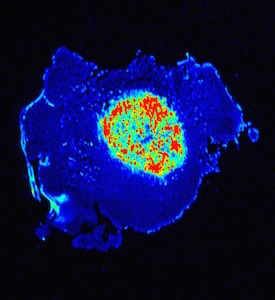


You must be logged in to post a comment.